. 2023 Jan 20;24(3):2121.
doi: 10.3390/ijms24032121.

Properties and Printability of the Synthesized Hydrogel Based on GelMA

Affiliations

Properties and Printability of the Synthesized Hydrogel Based on GelMA

Nadezhda V Arguchinskaya et al. Int J Mol Sci. .

Abstract

Gelatin methacryloyl (GelMA) has recently attracted increasing attention. Unlike other hydrogels, it allows for the adjustment of the mechanical properties using such factors as degree of functionalization, concentration, and photocrosslinking parameters. In this study, GelMA with a high degree of substitution (82.75 ± 7.09%) was synthesized, and its suitability for extrusion printing, cytocompatibility, and biocompatibility was studied. Satisfactory printing quality was demonstrated with the 15% concentration hydrogel. The high degree of functionalization led to a decrease in the ability of human adipose-derived stem cells (ADSCs) to adhere to the GelMA surface. During the first 3 days after sowing, proliferation was observed. Degradation in animals after subcutaneous implantation was slowed down.

Keywords: GelMA; biocompatibility; cytocompatibility; extrusion 3D bioprinting; hydrogel.


Conflict of interest statement

The authors declare no conflict of interest.

Figures

Figure 1
Figure 1
Ninhydrin assay. An example of coloring gelatin at a concentration of 1–10 mg/mL to obtain a standard curve using ninhydrin analysis (A). The value of the optical density of gelatin from 1–10 mg/mL (B). The linear section of the standard curve, which was used to build a linear approximation (C).
Figure 2
Figure 2
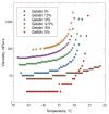
Dependence of the viscosity of hydrogels GelMA 15% and gelatin 5%, 7.5%, 10%, 12.5%, and 15% on temperature.
Figure 3
Figure 3
Printability of synthesized hydrogel. Formation of a 15% GelMA filament during extrusion through a 21G needle (A). The appearance of the maximum print speed estimation panel (B). The blue line marks the printed object, the red line marks the direction of ‘Skirt’ command printing at 1 mm/s, and the green line marks the direction of object printing at the speed range under study. The resulting panel (C). An example of an object printed from 15% GelMA before (D) and after a 72 h incubation (E). The appearance of the angled print panel (F) and the resulting panel at a speed of 5 mm/s (G) and 1 mm/s (H).
Figure 4
Figure 4
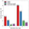
Proliferation of ADSCs in DMEM without gelatin, in the presence of unmodified porcine skin gelatin (type A, Bloom 300), synthesized, and commercialized by GelMA. For each group, the number of cells (X¯±SX¯) is shown on the 3rd and 7th days after seeding.
Figure 5
Figure 5
Implant from the synthesized GelMA in the subcutaneous tissue of rats after 10 days (indicated by the letters Sc). The capsule is shown by arrows. Staining with hematoxylin and eosin. Magnification of the image—×5 (A). Fragments of an implant in a connective tissue capsule (indicated by the letters Sc) surrounded by multinucleated macrophages (shown by arrows). Staining with hematoxylin and eosin. Magnification of the image—×20 (B).
Figure 6
Figure 6
Implant from synthesized GelMA in the subcutaneous tissue of rats after 17 days (indicated by the letters Sc). Connective tissue capsule around the implant (shown with asterisks). Multinucleated macrophages destroying scaffold fragments (shown by arrows). Staining with hematoxylin and eosin. Magnification of the image—×10 (A). Enlarged fragment. Arrows show multinucleated scaffold resorption cells. Staining with hematoxylin and eosin. Magnification of the image—×20 (B).
Figure 7
Figure 7
Implant from synthesized GelMA in the subcutaneous tissue of rats after 33 days (marked with Sc), surrounded by a connective tissue capsule. Connective tissue capsule around the implant (shown by arrows). Staining with hematoxylin and eosin. Magnification of the image—×20 (A). Collagen fibers in the connective tissue capsule. Staining with hematoxylin and eosin. Magnification of the image—×40 (B). Connective tissue of the capsule around the implant. Staining with hematoxylin and eosin. Magnification of the image—×40 (C). Fragments of the implant. Staining with hematoxylin and eosin. Magnification of the image—×10 (D). Magnification of the image—×40. Arrows show multinucleated macrophages (E). Circumcellular infiltrate (marked with arrows) in the border zone between the capsule and the implant (marked with Sc) after 33 days. Hematoxylin and eosin staining. Magnification of the image—×40 (F).
Figure 8
Figure 8
Scaffold preparation for in vivo implantation. Photograph of the printed PLA form (A). Scaffolds obtained by molding (B).

References

    1. Leberfinger A.N., Dinda S., Wu Y., Koduru S.V., Ozbolat V., Ravnic D.J., Ozbolat I.T. Bioprinting functional tissues. Acta Biomater. 2019;95:32–49. doi: 10.1016/j.actbio.2019.01.009. - DOI - PMC - PubMed
    1. Fan C., Wang D.-A. Macroporous hydrogel scaffolds for three-dimensional cell culture and tissue engineering. Tissue Eng. Part B Rev. 2017;23:451–461. doi: 10.1089/ten.teb.2016.0465. - DOI - PubMed
    1. Annabi N., Tamayol A., Uquillas J.A., Akbari M., Bertassoni L.E., Cha C., Camci-Unal G., Dokmeci M.R., Peppas N.A., Khademhosseini A. 25th Anniversary Article: Rational Design and Applications of Hydrogels in Regenerative Medicine. Adv. Mater. 2014;26:85–124. doi: 10.1002/adma.201303233. - DOI - PMC - PubMed
    1. Isaeva E.V., Beketov E.E., Demyashkin G.A., Yakovleva N.D., Arguchinskaya N.V., Kisel A.A., Lagoda T.S., Malakhov E.P., Smirnova A.N., Petriev V.M., et al. Cartilage Formation In Vivo Using High Concentration Collagen-Based Bioink with MSC and Decellularized ECM Granules. Int. J. Mol. Sci. 2022;23:2703. doi: 10.3390/ijms23052703. - DOI - PMC - PubMed
    1. Gao Q., Kim B.S., Gao G. Advanced Strategies for 3D Bioprinting of Tissue and Organ Analogs Using Alginate Hydrogel Bioinks. Mar. Drugs. 2021;19:708. doi: 10.3390/md19120708. - DOI - PMC - PubMed

LinkOut - more resources