Review
. 2023 Jan 22;24(3):2202.
doi: 10.3390/ijms24032202.

The Cell-Specific Role of SHP2 in Regulating Bone Homeostasis and Regeneration Niches

Affiliations
Review

The Cell-Specific Role of SHP2 in Regulating Bone Homeostasis and Regeneration Niches

Jie Zhang et al. Int J Mol Sci. .

Abstract

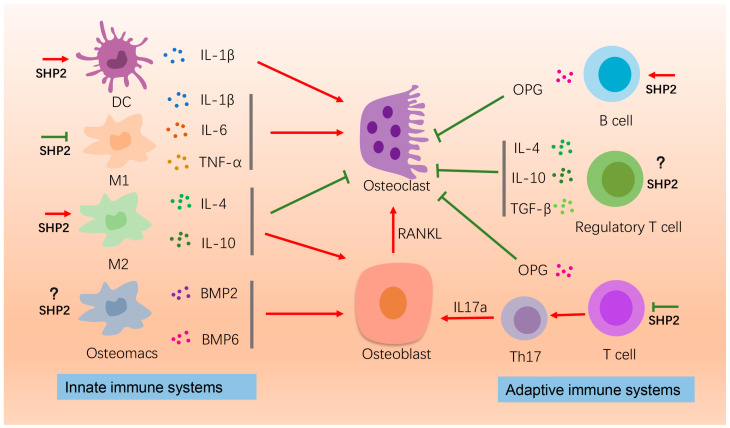
Src homology-2 containing protein tyrosine phosphatase (SHP2), encoded by PTPN11, has been proven to participate in bone-related diseases, such as Noonan syndrome (NS), metachondromatosis and osteoarthritis. However, the mechanisms of SHP2 in bone remodeling and homeostasis maintenance are complex and undemonstrated. The abnormal expression of SHP2 can influence the differentiation and maturation of osteoblasts, osteoclasts and chondrocytes. Meanwhile, SHP2 mutations can act on the immune system, vasculature and nervous system, which in turn affect bone development and remodeling. Signaling pathways regulated by SHP2, such as mitogen-activated protein kinase (MAPK), Indian hedgehog (IHH) and phosphatidylinositol-4,5-bisphosphate 3-kinase (PI3K)/protein kinase B (AKT), are also involved in the proliferation, differentiation and migration of bone functioning cells. This review summarizes the recent advances of SHP2 on osteogenesis-related cells and niche cells in the bone marrow microenvironment. The phenotypic features of SHP2 conditional knockout mice and underlying mechanisms are discussed. The prospective applications of the current agonists or inhibitors that target SHP2 in bone-related diseases are also described. Full clarification of the role of SHP2 in bone remodeling will shed new light on potential treatment for bone related diseases.

Keywords: SHP2; SHP2 agonist; SHP2 inhibitor; bone microenvironment; bone remodeling; homeostasis.


Conflict of interest statement

The authors declare no conflict of interest. The funders had no role in the design of the study; in the collection, analyses, or interpretation of data; in the writing of the manuscript; or in the decision to publish the results.

Figures

Figure 1
Figure 1
The effects of SHP2 on bone homeostasis by affecting immune cells. Here the roles of macrophages, B-cells, and T-cells on osteogenesis or osteoclatogenesis are depicted. IL-1β, IL-6, TNF-α released by the M1 macrophages promote osteoclastogenesis for bone resorption. SHP2 suppresses this process. The M2 macrophages secrete IL-4 and IL-10 to promote bone formation and suppress bone resorption. SHP2 enhances this process. Osteomacs produce BMP2 and BMP6 to promote bone formation. The effect of SHP2 on osteomacs is unknown. B-cells produce OPG to inhibit osteoclastogenesis and SHP2 promotes pre-B cells proliferation. Regulatory T cell releases IL-4, IL-10 and TNF-α to inhibit osteoclastogenesis. T cells, on the one hand, produce OPG to inhibit osteoclasts, and on the other hand differentiate into TH17, promoting osteoblast activity by producing IL-17α. This process is inhibited by SHP2. “Red arrow” refers to promotion; “Green arrow” refers to inhibition.
Figure 2
Figure 2
The effects of SHP2 on bone homeostasis by affecting vasculature ECs. In physiological situations, ECs release multiple cytokines, such as HIF-1α, Noggin, FGF1, TGFβ, etc., to promote osteogenesis. In pathological conditions, such as in inflammatory conditions, ECs secrete MMP9 and RANKL to promote osteoclastogenesis. Deletion of SHP2 in ECs decreases HIF-1α expression. Furthermore, the junctions between ECs are disordered and the integrity of the vascular barrier is disrupted. The SHP2-deficient ECs undergo apoptosis through activation of MAPK and AKT pathways. “Red arrow” refers to promotion.
Figure 3
Figure 3
The role of the nervous system on osteoblasts and osteoclasts. In peripheral nervous system, sensory and sympathetic nerves secrete neurotransmitters to regulate bone formation and resorption. CGRP suppresses osteoclasts formation. CGRP and SP promotes osteogenesis via increasing BMP2 and RUNX2 expression. VIP promotes osteogenesis through WNT/β-catenin signaling. In central nervous system, neurons can release extracellular vesicles enriched with miR-328a-3P and miR-150-5P to promote osteogenesis. “Red arrow” refers to promotion; “Green arrow” refers to inhibition.

Similar articles

Cited by

References

    1. Sommerfeldt D.W., Rubin C.T. Biology of bone and how it orchestrates the form and function of the skeleton. Eur. Spine J. 2001;10:S86–S95. doi: 10.1007/s005860100283. - DOI - PMC - PubMed
    1. Salhotra A., Shah H.N., Levi B., Longaker M.T. Mechanisms of bone development and repair. Nat. Rev. Mol. Cell Biol. 2020;21:696–711. doi: 10.1038/s41580-020-00279-w. - DOI - PMC - PubMed
    1. Berendsen A.D., Olsen B.R. Bone development. Bone. 2015;80:14–18. doi: 10.1016/j.bone.2015.04.035. - DOI - PMC - PubMed
    1. Lopes D., Martins-Cruz C., Oliveira M.B., Mano J.F. Bone physiology as inspiration for tissue regenerative therapies. Biomaterials. 2018;185:240–275. doi: 10.1016/j.biomaterials.2018.09.028. - DOI - PMC - PubMed
    1. Qin Q., Lee S., Patel N., Walden K., Gomez-Salazar M., Levi B., James A.W. Neurovascular coupling in bone regeneration. Exp. Mol. Med. 2022;54:1844–1849. doi: 10.1038/s12276-022-00899-6. - DOI - PMC - PubMed

MeSH terms

Substances

LinkOut - more resources