. 2023 Jan 24;24(3):2295.
doi: 10.3390/ijms24032295.

Role of Histone Tails and Single Strand DNA Breaks in Nucleosomal Arrest of RNA Polymerase

Affiliations

Role of Histone Tails and Single Strand DNA Breaks in Nucleosomal Arrest of RNA Polymerase

Nadezhda S Gerasimova et al. Int J Mol Sci. .

Abstract

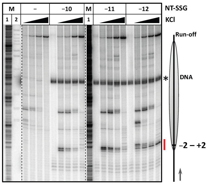
Transcription through nucleosomes by RNA polymerases (RNAP) is accompanied by formation of small intranucleosomal DNA loops (i-loops). The i-loops form more efficiently in the presence of single-strand breaks or gaps in a non-template DNA strand (NT-SSBs) and induce arrest of transcribing RNAP, thus allowing detection of NT-SSBs by the enzyme. Here we examined the role of histone tails and extranucleosomal NT-SSBs in i-loop formation and arrest of RNAP during transcription of promoter-proximal region of nucleosomal DNA. NT-SSBs present in linker DNA induce arrest of RNAP +1 to +15 bp in the nucleosome, suggesting formation of the i-loops; the arrest is more efficient in the presence of the histone tails. Consistently, DNA footprinting reveals formation of an i-loop after stalling RNAP at the position +2 and backtracking to position +1. The data suggest that histone tails and NT-SSBs present in linker DNA strongly facilitate formation of the i-loops during transcription through the promoter-proximal region of nucleosomal DNA.

Keywords: DNA damage; DNA loop; chromatin structure; nucleosome; single-strand DNA breaks; transcription-coupled DNA repair.


Conflict of interest statement

The authors declare no conflict of interest.

Figures

Figure 1
Figure 1
Experimental strategy for analysis of the effect of NT-SSBs on transcription through chromatin. (A) A model describing the effect of NT-SSBs on transcription through a nucleosome [27,29,30]. As RNAP encounters a nucleosome (intermediate 1), transient intranucleosomal i-loops can be formed (intermediate 2). An NT-SSB positioned behind the transcribing enzyme (shown by red star) greatly stabilizes the i-loop, inducing arrest of RNAP (intermediate 3) [30]. Direction of transcription is indicated by green and black arrows. (B) The 603 nucleosomes containing unique ssDNA gap (−12, −11 or −10) or break (+2) in the non-template strand are assembled on the DNA and transcribed for different time intervals in the presence of various concentrations of KCl. Pulse-labeled RNA (the label is shown by black asterisk) is separated by denaturing PAGE. Non-template DNA strand is 5’-labeled (shown by the black stars). Transcription start site is indicated by arrow. (C) An example of nucleosomes with ssDNA gap at the position −11 bp in nucleosomal DNA assembled using histone octamers with or without histone tails. DNA was radioactively labeled at 5′-end. Analysis by non-denaturing PAGE.
Figure 2
Figure 2
NT-SSB+2 induces arrest of RNAP during transcription of the +(10–20) region. (A) Transcription through 603 nucleosomes with and without single ssDNA break at the position +2 by E. coli RNAP for 2 min at 40, 150 or 300 mM KCl. Analysis of pulse-labeled RNA by denaturing PAGE. M—pBR322 MspI digest. Position of the nucleosome and direction of transcription are shown by the grey oval and arrow on the right, respectively. The +(10–20) region of RNAP arrest is shown by the red line. (B) Quantitative analysis of the RNA products obtained after transcription of the template with and without a nick at the position +2 at 40 mM KCl (averages of 3 experiments and standard deviations are shown). (C) The expected structure if the i-loop formed during transcription of the +(10–15) region. NT-SSB+2 is indicated by red star.
Figure 3
Figure 3
Single strand gaps at positions −10, −11 and −12 induce arrest of RNAP at the nucleosome entry site. Transcription of 603 nucleosomes containing single NT-SSGs at the positions −12, −11 or −10 by E. coli RNAP for 5 min at 40, 150, 300 or 1000 mM KCl. Analysis of pulse-labeled RNA by denaturing PAGE. M1—RNA marker (see Methods). M2—pBR322 MspI digest. The radioactively labeled fragment of the gap-containing non-template DNA strand (103 nucleotides in length) is shown by the asterisk. The region of RNAP arrest near position +1 in presence of NT-SSGs is shown by the red line. Other designations as in Figure 2A.
Figure 4
Figure 4
Removal of histone tails results in relief of nick- and gap-induced arrest of RNAP at +(1–5) and +(13–19) regions. (A) Nucleosomes NT-SSB+2 with and without histone tails were transcribed by E. coli RNAP for 2 min at 40 mM KCl. Analysis of pulse-labeled RNA by denaturing PAGE. M—pBR322 MspI digest. The regions of RNAP pausing/arrest affected by the presence of histone tails are indicated by red lines. (B) Nucleosomes with and without histone tails with NT-SSG at the position −11 were transcribed by E. coli RNAP for 5 min at 40, 150, 300 or 1000 mM KCl. Analysis of pulse-labeled RNA by denaturing PAGE. M—pBR322 MspI digest. The asterisks indicate radioactively labeled fragments of the DNA template (103 and 273 nucleotides long). Position of the nucleosome and direction of transcription are shown by the grey oval and arrow on the right, respectively. (C) Time courses of transcription through the nucleosomes (intact and with NT-SSG-11) with and without tails. The templates were transcribed by E. coli RNAP for 1 sec, 2 sec, 4 sec, 8 sec, 1 min, 5 min or 10 min at 150 mM KCl. Analysis of the pulse-labeled RNA by denaturing PAGE. M—pBR322 MspI digest. Radioactively labeled fragments of the DNA template (103 and 273 nucleotides in length) are shown by asterisks. Position of the nucleosome and direction of transcription are shown by the grey oval and arrow, respectively. The region of RNAP pausing near the position +1 in presence of ss-gap is indicated by the red line.
Figure 5
Figure 5
Histone tails strongly contribute to protection of nucleosomal and linker DNA from hydroxyl radicals. The 5′-end of the non-template DNA strand was radioactively labeled. Nucleosomes with or without histone tails were incubated in the presence of hydroxyl radicals. End-labeled DNA was analyzed by denaturing PAGE (on the left). M—pBR322 MspI digest. Lanes labeled by colored asterisks (black for markers, green for nucleosomes with histone tails and red for nucleosomes without histone tails) are also shown at a higher magnification in the middle; corresponding scans are shown on the right. Nucleosome position is shown by the grey oval. Red lines and arrows indicate DNA regions protected from hydroxyl radicals by the histone tails.
Figure 6
Figure 6
Formation of an i-loop containing backtracked RNAP at the position +1. (A) On the left: EC-5 and EC+2 were formed on the 603 DNA and nucleosomes and incubated in the presence of hydroxyl radicals. End-labeled DNA was analyzed by denaturing PAGE (on the left). M—pBR322 MspI digest. Nucleosome position is indicated by grey oval. On the right: Scans of the lanes with nucleosomes and nucleosomal ECs. Purple and blue lines indicate DNA regions protected from hydroxyl radicals by RNA polymerase and DNA region upstream the RNAP protected only in EC+2, respectively. (B) EC+2 contains an unstable i-loop. Direction of transcription and arrest of RNAP are indicated by black arrows and red cross, respectively. (C) Incubation of EC+2 in the presence of 10, 20 or 40 nM of GreB. Analysis of pulse-labeled RNA by denaturing PAGE. M—pBR322 MspI digest. Positions of active center of RNAP are indicated on the right.
Figure 7
Figure 7
Proposed roles of NT-SSBs and histone N-terminal tails during transcription of the early region of nucleosomal DNA containing a damage. Transcription of the linker and early nucleosomal DNA (intermediate 1) induces transient DNA uncoiling from the histone octamer (intermediate 2) and formation of an unstable intranucleosomal i-loops when the active center of RNA polymerase traverses the −3 to +19 bp DNA region (intermediate 3); the i-loops often incorporate linker DNA. I-loops are stabilized by NT-SSBs (indicated by red stars) and by the histone tails. Histone modifications of the tails and factors interacting with core histones or RNA polymerase could affect the efficiency of loop formation.

Similar articles

Cited by

References

    1. Tubbs A., Nussenzweig A. Endogenous DNA Damage as a Source of Genomic Instability in Cancer. Cell. 2017;168:644–656. doi: 10.1016/j.cell.2017.01.002. - DOI - PMC - PubMed
    1. Lindahl T. Instability and decay of the primary structure of DNA. Nature. 1993;362:709–715. doi: 10.1038/362709a0. - DOI - PubMed
    1. Pogozelski W.K., Tullius T.D. Oxidative Strand Scission of Nucleic Acids: Routes Initiated by Hydrogen Abstraction from the Sugar Moiety. Chem. Rev. 1998;98:1089–1108. doi: 10.1021/cr960437i. - DOI - PubMed
    1. Caldecott K.W. DNA single-strand break repair and human genetic disease. Trends Cell Biol. 2022;32:733–745. doi: 10.1016/j.tcb.2022.04.010. - DOI - PubMed
    1. Caldecott K.W., Ward M.E., Nussenzweig A. The threat of programmed DNA damage to neuronal genome integrity and plasticity. Nat. Genet. 2022;54:115–120. doi: 10.1038/s41588-021-01001-y. - DOI - PubMed

MeSH terms