. 2023 Jan 25;24(3):2372.
doi: 10.3390/ijms24032372.

Epigenetic Immune Cell Counting to Analyze Potential Biomarkers in Preterm Infants: A Proof of Principle in Necrotizing Enterocolitis

Affiliations

Epigenetic Immune Cell Counting to Analyze Potential Biomarkers in Preterm Infants: A Proof of Principle in Necrotizing Enterocolitis

Michiel H D Schoenaker et al. Int J Mol Sci. .

Abstract

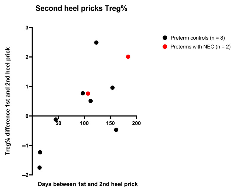
Epigenetic immune cell counting is a DNA (de)methylation-based technique which can be used to quantify lymphocyte subsets on dried blood spots (DBS). The foregoing techniques allow for a retrospective investigation of immune cell profiles in newborns. In this study, we used this technique for determining lymphocyte subcounts as a potential biomarker for necrotizing enterocolitis (NEC). We investigated whether this technique can be implemented in the field of neonatology, by testing whether regulatory T cell (Treg) levels are pre-existently low in preterms with NEC. Newborn screening (NBS) cards from 32 preterms with NEC and 32 age- and weight-matched preterm controls, and 60 healthy term newborns, were analyzed. Relative and absolute cell counts were determined for CD3+, CD4+, CD8+, Th17, and Treg T cells. For both relative and absolute cell counts of CD3+, CD4+, CD8+, and Th17 T cells, significant differences were found between healthy term controls and both preterm groups, but not between preterm groups. For Tregs, no significant differences were found in either relative or absolute counts between any of the newborn groups. This study demonstrates the principle of epigenetic immune cell counting to analyze lymphocyte subsets in preterm neonates.

Keywords: Th17 cell; epigenetic immune cell counting; necrotizing enterocolitis; neonate; preterm; regulatory T cell.


Conflict of interest statement

JW is an employee of the company Epimune GmbH. The other authors declare no competing interest.

Figures

Figure 1
Figure 1
The principle of epigenetic immune cell counting by amplification of cell type-specific demethylated genomic regions, adapted from Epimune GmbH.
Figure 2
Figure 2
(AE) Relative (epi) CD3+, CD4+, CD8+, Th17, and Treg T cell counts (% of demethylated gene copies of total GAPDH demethylated copies) of healthy term controls (n = 60), preterm controls (n = 32) and preterms with NEC (n = 32), presented on the X-axis. Y-axis displays the relative cell count as percentage of total leukocytes. (F) Relative Treg/CD4 ratio. Groups are presented on the X-axis, with the Treg/CD4+ ratio on the Y-axis. Pairwise significances are represented at the top of the graph, with ns meaning non-significant, ** meaning p < 0.01, *** meaning p < 0.001. Black lines indicate the median value.
Figure 3
Figure 3
(AE) Estimated absolute CD3+, CD4+, CD8+, Th17, and Treg T cell counts measured in dried blood spots of healthy term controls (n = 60), preterm controls (n = 28), and preterms with NEC (n = 28), presented on the X-axis. Y-axis depicts the number of cells per µL. Pairwise significances are represented at the top of the graph, with ns meaning non-significant, * meaning p < 0.05, ** meaning p < 0.01, *** meaning p < 0.001.
Figure 3
Figure 3
(AE) Estimated absolute CD3+, CD4+, CD8+, Th17, and Treg T cell counts measured in dried blood spots of healthy term controls (n = 60), preterm controls (n = 28), and preterms with NEC (n = 28), presented on the X-axis. Y-axis depicts the number of cells per µL. Pairwise significances are represented at the top of the graph, with ns meaning non-significant, * meaning p < 0.05, ** meaning p < 0.01, *** meaning p < 0.001.
Figure 4
Figure 4
Different stages of NEC and relative Treg cell counts. The X-axis displays the NEC stage, while the Y-axis shows the relative Treg cell count. Black lines represent the median value per group.
Figure 5
Figure 5
The number of days between the first and second heel prick (X-axis) and the difference in relative Treg cell counts between the heel pricks (Y-axis). Preterms with NEC are indicated in red (n = 2). Preterm controls are indicated in black (n = 8).

References

    1. Kleen T.O., Yuan J. Quantitative Real-Time PCR Assisted Cell Counting (QPACC) for Epigenetic—Based Immune Cell Quantification in Blood and Tissue. J. Immunother Cancer. 2015;3:46. doi: 10.1186/s40425-015-0087-8. - DOI - PMC - PubMed
    1. Baron U., Werner J., Schildknecht K., Schulze J.J., Mulu A., Liebert U.-G., Sack U., Speckmann C., Gossen M., Wong R.J., et al. Epigenetic Immune Cell Counting in Human Blood Samples for Immunodiagnostics. Sci. Transl. Med. 2018;10:eaan3508. doi: 10.1126/scitranslmed.aan3508. - DOI - PubMed
    1. Bazacliu C., Neu J. Necrotizing Enterocolitis: Long Term Complications. Curr. Pediatr. Rev. 2019;15:115–124. doi: 10.2174/1573396315666190312093119. - DOI - PubMed
    1. Robinson J.R., Rellinger E.J., Hatch L.D., Weitkamp J.H., Speck K.E., Danko M., Blakely M.L. Surgical Necrotizing Enterocolitis. Semin. Perinatol. 2017;41:70–79. doi: 10.1053/j.semperi.2016.09.020. - DOI - PMC - PubMed
    1. Alganabi M., Lee C., Bindi E., Li B., Pierro A. Recent Advances in Understanding Necrotizing Enterocolitis [Version 1; Referees: 2 Approved] F1000Res. 2019;8:F1000 Faculty Rev-107. doi: 10.12688/f1000research.17228.1. - DOI - PMC - PubMed

Grants and funding