Review
. 2023 Jan 29;24(3):2555.
doi: 10.3390/ijms24032555.

The Significance of Cancer Stem Cells and Epithelial-Mesenchymal Transition in Metastasis and Anti-Cancer Therapy

Affiliations
Review

The Significance of Cancer Stem Cells and Epithelial-Mesenchymal Transition in Metastasis and Anti-Cancer Therapy

Lili Liang et al. Int J Mol Sci. .

Abstract

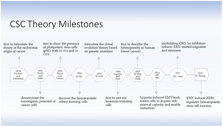
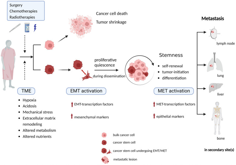
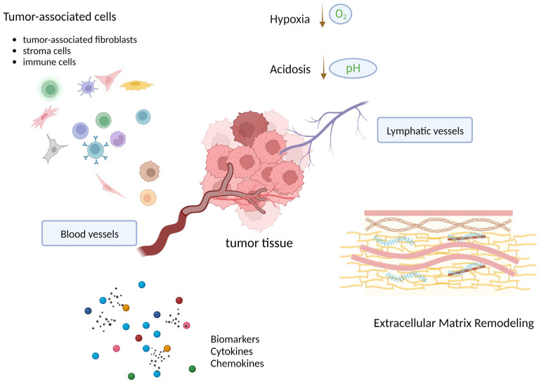
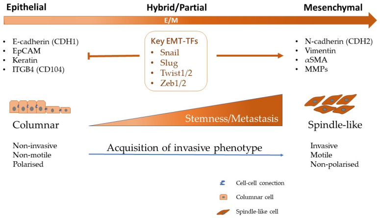
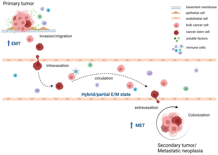
Cancer stem cells (CSCs) have been identified and characterized in both hematopoietic and solid tumors. Their existence was first predicted by Virchow and Cohnheim in the 1870s. Later, many studies showed that CSCs can be identified and isolated by their expression of specific cell markers. The significance of CSCs with respect to tumor biology and anti-cancer treatment lies in their ability to maintain quiescence with very slow proliferation, indefinite self-renewal, differentiation, and trans-differentiation such as epithelial-mesenchymal transition (EMT) and its reverse process mesenchymal-epithelial transition (MET). The ability for detachment, migration, extra- and intravasation, invasion and thereby of completing all necessary steps of the metastatic cascade highlights their significance for metastasis. CSCs comprise the cancer cell populations responsible for tumor growth, resistance to therapies and cancer metastasis. In this review, the history of the CSC theory, their identification and characterization and their biology are described. The contribution of the CSC ability to undergo EMT for cancer metastasis is discussed. Recently, novel strategies for drug development have focused on the elimination of the CSCs specifically. The unique functional and molecular properties of CSCs are discussed as possible therapeutic vulnerabilities for the development of novel anti-metastasis treatments. Prospectively, this may provide precise personalized anti-cancer treatments with improved therapeutic efficiency with fewer side effects and leading to better prognosis.

Keywords: epithelial–mesenchymal transition (EMT); hybrid EMT; invasion; mesenchymal–epithelial transition (MET); partial EMT; stemness; tumor initiating cells; tumor microenvironment (TME).


Conflict of interest statement

The authors declare no conflict of interest.

Figures

Figure 1
Figure 1
The development of the CSC theory. The theory of SCs and CSCs was initially conceived one and a half centuries ago. Virchow and Cohnheim first proposed the embryonic origin of cancer in 1870s. It took more than half a century until Furth and Kahn successfully established a mouse leukemia model indicating the possibility of tumor initiation by a minority of cancer cells. Since then, the CSC theory has been under challenge until more knowledge was accumulated. Today, the theory of CSCs has been substantiated and is regarded as reliable. CSCs have been demonstrated to be closely associated with EMT and metastasis [4,5,6,8,9,10,13,16,17,18].
Figure 2
Figure 2
CSCs play a crucial role in metastasis due to their unique characteristics. Undergoing conventional standard anti-cancer therapies and the multi-directional influences from TME are cellular stress factors that CSCs are supposed to survive by activating an EMT program and stay in a quiescent state to keep themselves alive during dissemination and adjustment to different conditions in a new TME, being stimulated to proliferate when successfully adjusted to distant organs/sites. TME, tumor microenvironment; CSC, cancer stem cell; EMT, epithelial mesenchymal transition; MET, mesenchymal epithelial transition.
Figure 3
Figure 3
Interactions of CSCs within the TME. A tumor tissue is a complex composition of tumor cells and tumor-associated host tissue cells, along with blood and lymphatic vessels, molecules such as cytokines (colored circles), and acellular structures such as ECM. There are multi-directional interactions within the TME. These interactions alter that TME into a CSC supportive niche, which is tightly linked with the presence of hypoxia, acidosis, ECM remodeling, nutrients alterations and necrotic processes. The niche supports both the survival and maintenance of the CSCs by facilitating the promotion of stemness and regulation of dormancy.
Figure 4
Figure 4
The EMT and its associated transcription factors. The EMT program requires the activation of a variety of TFs, including Snail and Slug, Zeb1 and Zeb2, Twist1 and Twist2, FOXC2 and GSC. It leads to the decreased expression of epithelial markers such as E-cadherin and increase the expression of mesenchymal markers such as N-cadherin. Accordingly, the morphologic and functional changes occur correspondently during the EMT process. The EMT regulation mediated by TFs has been proposed as a central key factor in the acquisition of stemness and promotion of metastasis. E, epithelial; M, mesenchymal; TFs, transcription factors; αSMA, alpha smooth muscle actin; MMPs, matrix metallopeptidases.
Figure 5
Figure 5
The invasion–metastasis cascade during cancer dissemination. Cancer cells acquire the necessary capabilities to complete the invasion–metastasis cascade. Firstly, the cancer cells at the primary tumor site spread into the surrounding tissue and pass through the epithelial basement membrane. Next, those cells successfully get access and invade into lymphatic and/or blood vessels (intravasation). These cells have to maintain cell viability during traveling in the circulation. When arriving at distant site(s), those disseminated cancer cells migrate out from the vessels (extravasation) to colonize in the target organ. Upon the process, small cell clones or disseminated cancer cells (micro-metastasis) must survive and finally, adjust to the new microenvironment, proliferate and form macroscopic metastasis (macro-metastasis). E, epithelial; M, mesenchymal; EMT, epithelial–mesenchymal transition; MET, mesenchymal–epithelial transition.

References

    1. De Angelis M.L., Francescangeli F., Zeuner A. Breast Cancer Stem Cells as Drivers of Tumor Chemoresistance, Dormancy and Relapse: New Challenges and Therapeutic Opportunities. Cancers. 2019;11:1569. doi: 10.3390/cancers11101569. - DOI - PMC - PubMed
    1. Garg M. Emerging roles of epithelial-mesenchymal plasticity in invasion-metastasis cascade and therapy resistance. Cancer Metastasis Rev. 2022;41:131–145. doi: 10.1007/s10555-021-10003-5. - DOI - PubMed
    1. Lambert A.W., Weinberg R.A. Linking EMT programmes to normal and neoplastic epithelial stem cells. Nat. Rev. Cancer. 2021;21:325–338. doi: 10.1038/s41568-021-00332-6. - DOI - PubMed
    1. Capp J.-P. Cancer Stem Cells: From Historical Roots to a New Perspective. J. Oncol. 2019;2019:5189232. doi: 10.1155/2019/5189232. - DOI - PMC - PubMed
    1. Furth J., Kahn M.C., Breedis C. The Transmission of Leukemia of Mice with a Single Cell. Am. J. Cancer. 1937;31:276–282.