. 2023 Jan 30;24(3):2586.
doi: 10.3390/ijms24032586.

Activation of the RARα Attenuated CSF Hypersecretion to Inhibit Hydrocephalus Development via Regulating the MAFB/MSR1 Pathway

Affiliations

Activation of the RARα Attenuated CSF Hypersecretion to Inhibit Hydrocephalus Development via Regulating the MAFB/MSR1 Pathway

Hanhai Zeng et al. Int J Mol Sci. .

Abstract

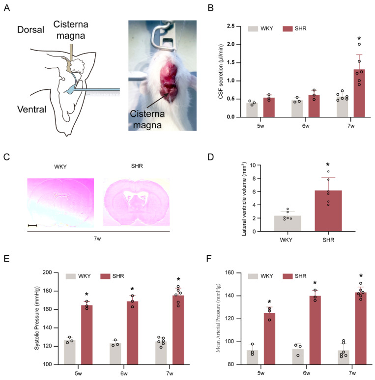
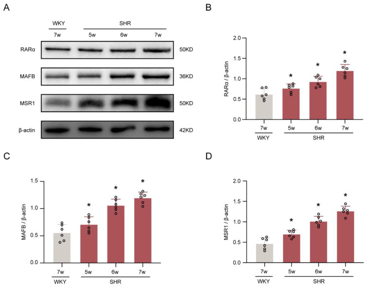
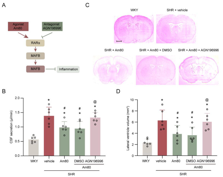
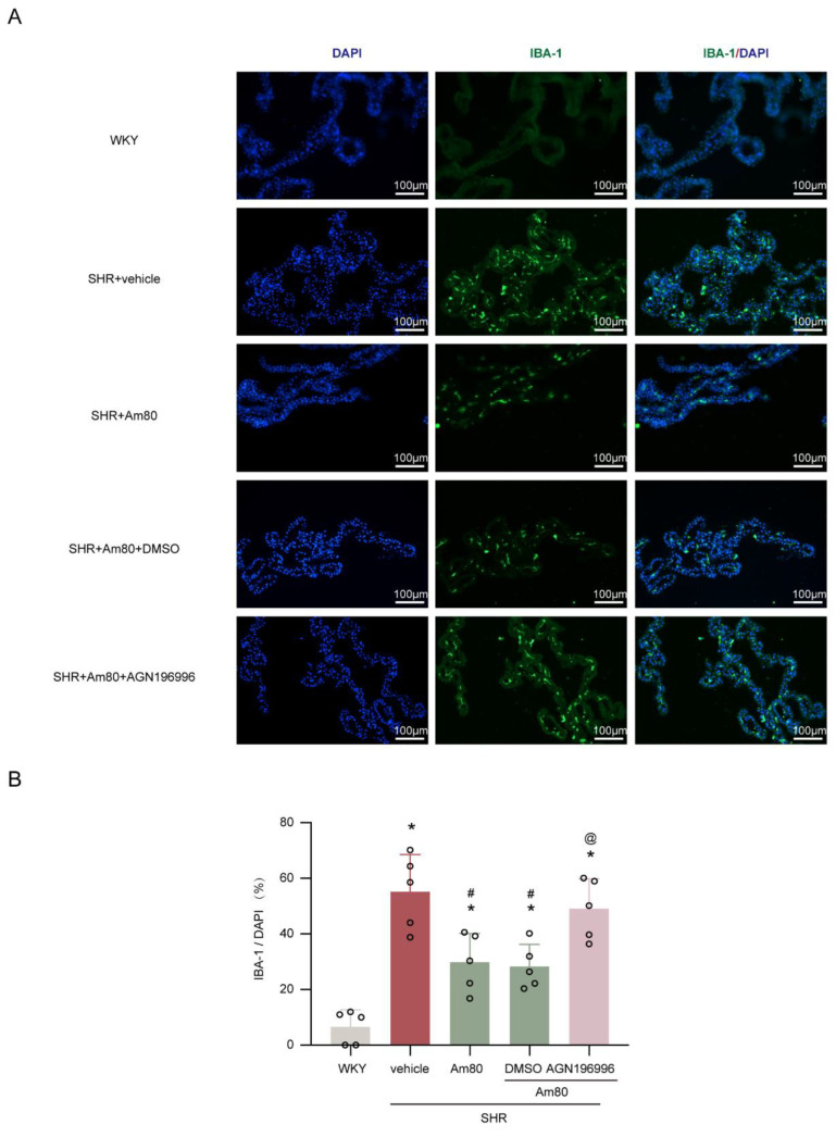
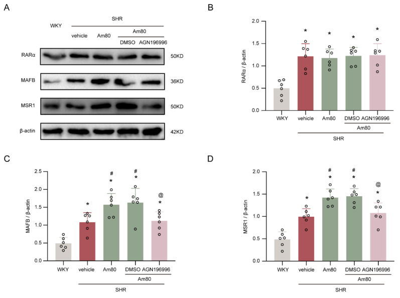
Hydrocephalus has been observed in rats with spontaneous hypertension (SHRs). It has been demonstrated that activation of the oxidative stress related protein retinoic acid receptor alpha (RARα) has neuroprotective impacts. Our investigation aims to determine the potential role and mechanism of RARα in hydrocephalus. The RARα-specific agonist (Am80) and RARα inhibitor (AGN196996) were used to investigate the role of RARα in cerebrospinal fluid (CSF) secretion in the choroid plexus of SHRs. Evaluations of CSF secretion, ventricular volume, Western blotting, and immunofluorescent staining were performed. Hydrocephalus and CSF hypersecretion were identified in SHRs but not in Wistar-Kyoto rats, occurring at the age of 7 weeks. The RARα/MAFB/MSR1 pathway was also activated in SHRs. Therapy with Am80 beginning in week 5 decreased CSF hypersecretion, hydrocephalus development, and pathological changes in choroid plexus alterations by week 7. AGN196996 abolished the effect of Am80. In conclusion, activation of the RARα attenuated CSF hypersecretion to inhibit hydrocephalus development via regulating the MAFB/MSR1 pathway. RARα may act as a possible therapeutic target for hydrocephalus.

Keywords: CSF; MSR1; RARα; hydrocephalus; spontaneous hypertensive rat.


Conflict of interest statement

The authors declare no conflict of interest.

Figures

Figure 1
Figure 1
CSF hypersecretion and hydrocephalus occurs in SHRs at seven weeks of age. (A) Left, schematic of the method used for in vivo quantification of CSF secretion in rats. Right, direct measurement of CSF secretion in a live rat. (B) Quantification of the rate of CSF secretion at weeks 5 (n = 3), 6 (n = 3), and 7 (n = 6). * p < 0.05 versus WKY rats at week 7. (C) Representative photomicrographs of coronal sections of rat brains (at −0.6 mm from the bregma) depicting ventricular volume at week 7 in SHRs compared with age-matched WKY rats. Scale bar, 2 mm. (D) Quantification of lateral ventricle volume in SHRs (n = 6) and WKY rats (n = 6) at weeks 7. * p < 0.05 versus WKY rats at week 7. (E,F) SHRs had significantly higher systolic blood pressure and mean arterial pressure than WKY rats at weeks 5 (n = 3), 6 (n = 3), and 7 (n = 6). * p < 0.05 versus WKY group. Values are means ± SD.
Figure 2
Figure 2
Temporal expression of RARα, MAFB, and MSR1 in SHRs. (A) Representative Western blotting images of RARα, MAFB, and MSR1. (BD) Quantitative analyses of RARα, MAFB, and MSR1. n = 6. * p < 0.05 versus WKY rats at week 7. Values are means ± SD.
Figure 3
Figure 3
AGN196996 abolished the protective effects of Am80. (A) Schematic diagram of the experimental design. (B) Quantification of the rate of CSF secretion at week 7 (n = 6). (C) Representative photomicrographs of coronal sections of rat brains (at −0.6 mm from the bregma), depicting ventricular volume at week 7. Scale bar, 2 mm. (D) Quantification of lateral ventricle volume at week 7 (n = 6). * p < 0.05 versus WKY rats at week 7, # p < 0.05 vs. SHR + vehicle group, @ p < 0.05 vs. SHR + Am80 + DMSO group. Values are means ± SD.
Figure 4
Figure 4
AGN196996 abolished the effects of Am80 on microglia/macrophage activation. (A) Examples of choroid plexus Iba-1 positive cells in rats at week 7. (B) Quantification of Iba-1 positive cells at week 7 (n = 5). Scale bar, 100 μm. * p < 0.05 versus WKY rats at week 7, # p < 0.05 vs. SHR + vehicle group, @ p < 0.05 vs. SHR + Am80 + DMSO group. Values are means ± SD.
Figure 5
Figure 5
Am80 stimulated RARα to reduce CSF hypersecretion via the MAFB/MSR1 pathway. (A) Representative Western blotting images of RARα, MAFB, and MSR1 at week 7. (BD) Quantitative analyses of RARα, MAFB, and MSR1 at week 7 (n = 6). * p < 0.05 versus WKY rats at week 7, # p < 0.05 vs. SHR + vehicle group, @ p < 0.05 vs. SHR + Am80 + DMSO group. Values are means ± SD.

Similar articles

Cited by

References

    1. Kahle K., Kulkarni A., Limbrick D., Warf B., Jr. Hydrocephalus in children. Lancet. 2016;387:788–799. - PubMed
    1. Strahle J., Garton H., Maher C., Muraszko K., Keep R., Xi G. Mechanisms of hydrocephalus after neonatal and adult intraventricular hemorrhage. Transl. Stroke Res. 2012;3((Suppl. S1)):25–38. - PMC - PubMed
    1. McAllister J., Williams M., 2nd, Walker M., Kestle J., Relkin N., Anderson A., Gross P., Browd S. An update on research priorities in hydrocephalus: Overview of the third National Institutes of Health-sponsored symposium “Opportunities for Hydrocephalus Research: Pathways to Better Outcomes”. J. Neurosurg. 2015;123:1427–1438. doi: 10.3171/2014.12.JNS132352. - DOI - PubMed
    1. Ritter S., Dinh T. Progressive postnatal dilation of brain ventricles in spontaneously hypertensive rats. Brain Res. 1986;370:327–332. doi: 10.1016/0006-8993(86)90488-9. - DOI - PubMed
    1. Leinonen V., Vanninen R., Rauramaa T. Cerebrospinal fluid circulation and hydrocephalus. Handb. Clin. Neurol. 2017;145:39–50. - PubMed