

Applying Metagenomic Analysis Using Nanopore Sequencer (MinION) for Precision Medicine in Bacterial Keratoconjunctivitis: Comprehensive Validation of Molecular Biological and Conventional Examinations
- PMID: 36768930
- PMCID: PMC9916690
- DOI: 10.3390/ijms24032611
Applying Metagenomic Analysis Using Nanopore Sequencer (MinION) for Precision Medicine in Bacterial Keratoconjunctivitis: Comprehensive Validation of Molecular Biological and Conventional Examinations
Abstract
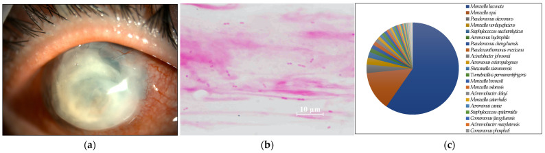
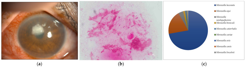
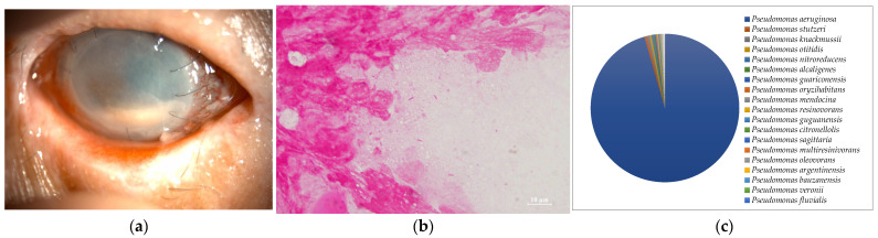
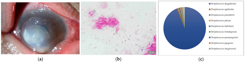
Smear microscopic examination and culture of the corneal scrapings are the gold standards for the diagnosis of bacterial keratoconjunctivitis. High-sensitivity molecular biological examinations of the ocular surface specimens are used clinically. However, the results require careful interpretation to avoid the unintentional detection of indigenous bacteria. Results of conventional and state-of-the-art examinations require clinical verification for specificity and sensitivity. In this study, smear microscopic examination, culture, and nanopore sequencing using the MinION of ocular surface specimens from eight clinically diagnosed bacterial keratoconjunctivitis cases were performed and compared. Seven of the eight cases (87.5%) were smear positive and five (62.5%) were culture positive. The former showed the same genus in >60% of the classified reads as one specific bacterium inferred from the smear microscopy when sequenced by the MinION. In two of the three culture-negative cases, the smear-positive images were highly reminiscent of the species comprising most of the MinION sequences. Four of the five culture-positive cases were consistent with the most prevalent bacteria in the sequencing results. Probable contamination among specimens processed on the same day were observed. In conclusion, the microscopic examination of the corneal scraping specimens may be more sensitive and specific than the culture examination. Additionally, although metagenomic analysis using the MinION contributes to more precise medication for bacterial keratoconjunctivitis, contamination can affect the results.
Keywords: bacterial keratoconjunctivitis; corneal scraping; culture; nanopore sequencer (MinION); smear microscopic examination.
Conflict of interest statement
The authors declare no conflict of interest.
Figures
Similar articles
-
Rapid identification of pathogens from positive blood culture bottles with the MinION nanopore sequencer.J Med Microbiol. 2018 Nov;67(11):1589-1595. doi: 10.1099/jmm.0.000855. Epub 2018 Oct 12. J Med Microbiol. 2018. PMID: 30311873
-
Real-time analysis of nanopore-based metagenomic sequencing from infected orthopaedic devices.BMC Genomics. 2018 Sep 27;19(1):714. doi: 10.1186/s12864-018-5094-y. BMC Genomics. 2018. PMID: 30261842 Free PMC article.
-
16S rRNA nanopore sequencing for the diagnosis of ocular infection: a feasibility study.BMJ Open Ophthalmol. 2022 May;7(1):e000910. doi: 10.1136/bmjophth-2021-000910. Epub 2022 May 24. BMJ Open Ophthalmol. 2022. PMID: 36161861 Free PMC article.
-
An identification protocol for ESBL-producing Gram-negative bacteria bloodstream infections using a MinION nanopore sequencer.J Med Microbiol. 2019 Aug;68(8):1219-1226. doi: 10.1099/jmm.0.001024. Epub 2019 Jun 25. J Med Microbiol. 2019. PMID: 31237534
-
On-Site MinION Sequencing.Adv Exp Med Biol. 2019;1129:143-150. doi: 10.1007/978-981-13-6037-4_10. Adv Exp Med Biol. 2019. PMID: 30968366 Review.
Cited by
-
Overview of Microorganisms: Bacterial Microbiome, Mycobiome, Virome Identified Using Next-Generation Sequencing, and Their Application to Ophthalmic Diseases.Microorganisms. 2025 Jun 3;13(6):1300. doi: 10.3390/microorganisms13061300. Microorganisms. 2025. PMID: 40572188 Free PMC article. Review.
References
-
- Flaxman S.R., Bourne R.R.A., Resnikoff S., Ackland P., Braithwaite T., Cicinelli M.V., Das A., Jonas J.B., Keeffe J., Kempen J.H., et al. Global causes of blindness and distance vision impairment 1990–2020: A systematic review and meta-analysis. Lancet Glob Health. 2017;5:e1221–e1234. doi: 10.1016/S2214-109X(17)30393-5. - DOI - PubMed
MeSH terms
LinkOut - more resources
Full Text Sources
