. 2023 Jan 31;24(3):2644.
doi: 10.3390/ijms24032644.

Clathrin-Mediated Albumin Clearance in Alveolar Epithelial Cells of Murine Precision-Cut Lung Slices

Affiliations

Clathrin-Mediated Albumin Clearance in Alveolar Epithelial Cells of Murine Precision-Cut Lung Slices

Vitalii Kryvenko et al. Int J Mol Sci. .

Abstract

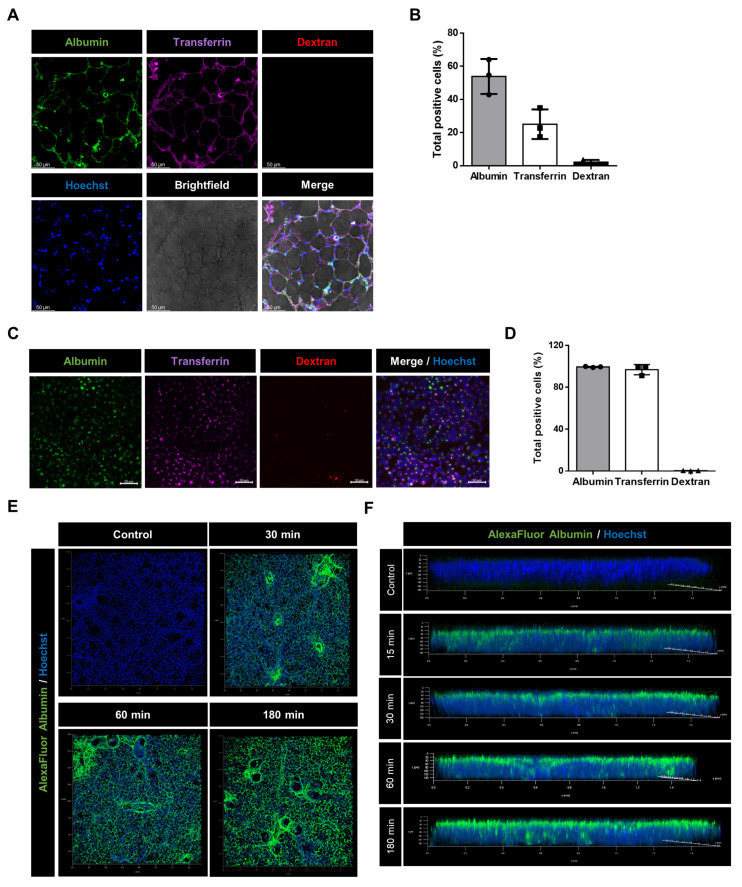
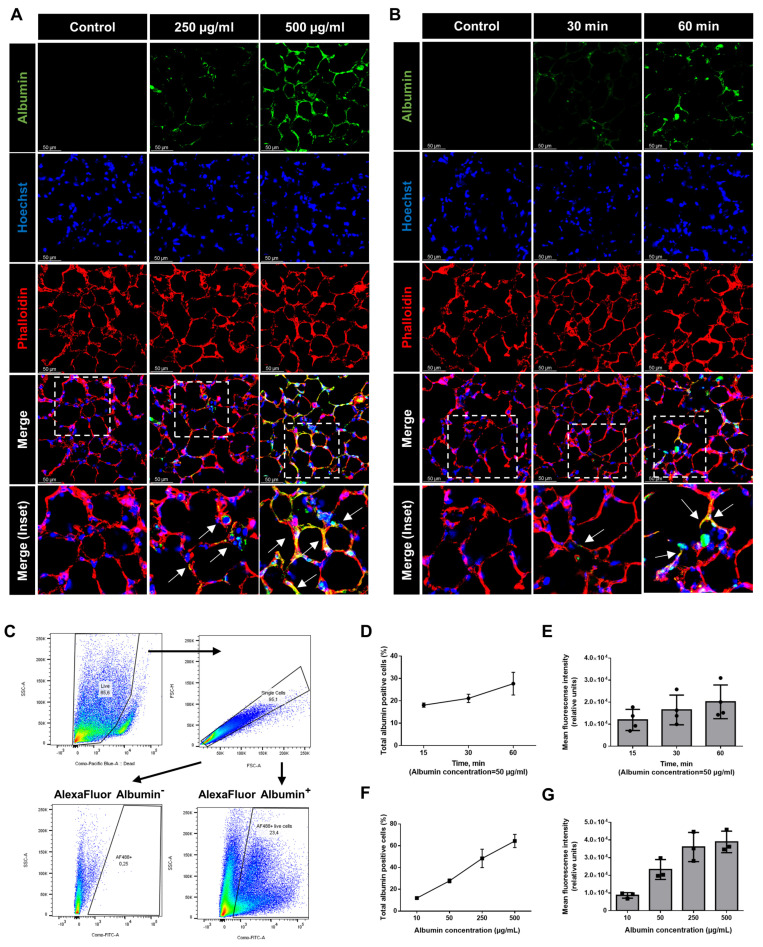
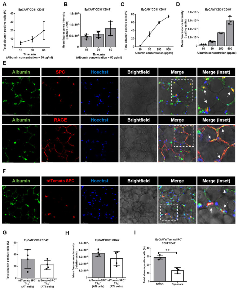
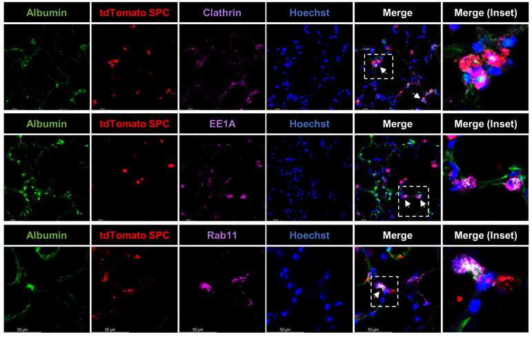
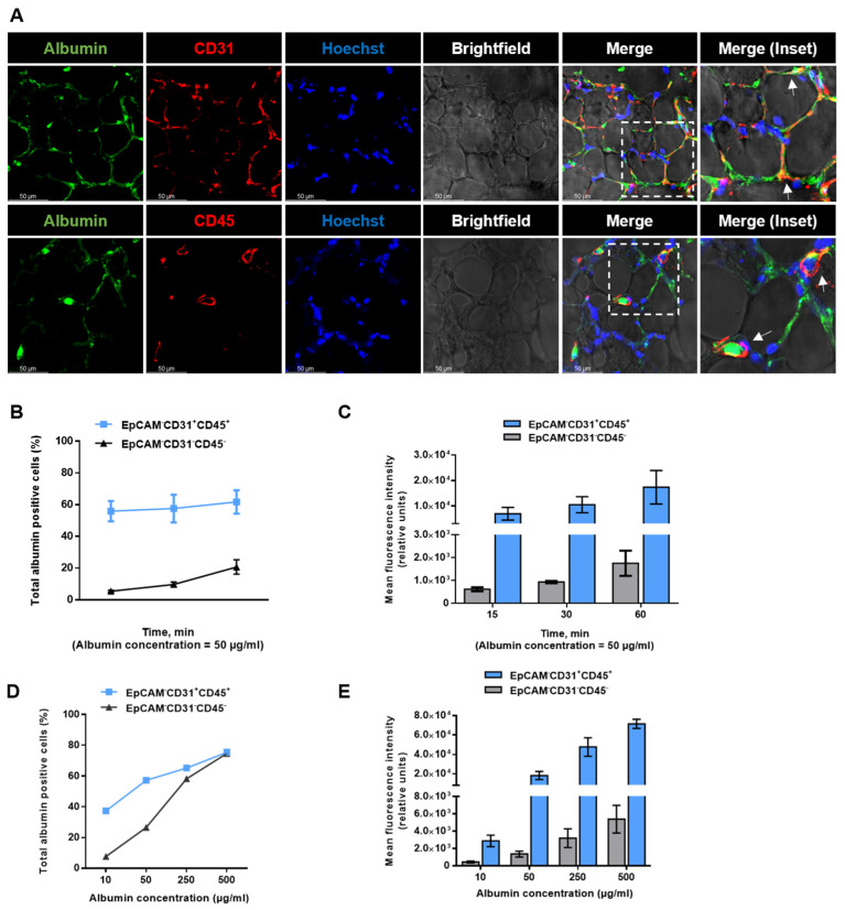
A hallmark of acute respiratory distress syndrome (ARDS) is an accumulation of protein-rich alveolar edema that impairs gas exchange and leads to worse outcomes. Thus, understanding the mechanisms of alveolar albumin clearance is of high clinical relevance. Here, we investigated the mechanisms of the cellular albumin uptake in a three-dimensional culture of precision-cut lung slices (PCLS). We found that up to 60% of PCLS cells incorporated labeled albumin in a time- and concentration-dependent manner, whereas virtually no uptake of labeled dextran was observed. Of note, at a low temperature (4 °C), saturating albumin receptors with unlabeled albumin and an inhibition of clathrin-mediated endocytosis markedly decreased the endocytic uptake of the labeled protein, implicating a receptor-driven internalization process. Importantly, uptake rates of albumin were comparable in alveolar epithelial type I (ATI) and type II (ATII) cells, as assessed in PCLS from a SftpcCreERT2/+: tdTomatoflox/flox mouse strain (defined as EpCAM+CD31-CD45-tdTomatoSPC-T1α+ for ATI and EpCAM+CD31-CD45-tdTomatoSPC+T1α- for ATII cells). Once internalized, albumin was found in the early and recycling endosomes of the alveolar epithelium as well as in endothelial, mesenchymal, and hematopoietic cell populations, which might indicate transcytosis of the protein. In summary, we characterize albumin uptake in alveolar epithelial cells in the complex setting of PCLS. These findings may open new possibilities for pulmonary drug delivery that may improve the outcomes for patients with respiratory failure.

Keywords: acute respiratory distress syndrome; albumin; alveolar epithelium; endocytosis; precision-cut lung slices; protein transport.


Conflict of interest statement

The authors declare no conflict of interest.

Figures

Figure 1
Figure 1
Albumin, transferrin, and dextran uptake in murine PCLS and mice MLE-12 epithelial cells: (A) PCLS were incubated for 1 h in a solution containing 250 μg/mL of AlexaFluor488-albumin (green), AlexaFluor647-transferrin (magenta), and lysine-fixable TexasRed-dextran (70,000 Da and red). Confocal microscopy images are shown. Scale bar−50 µM. (B) Flow cytometry analysis showing frequency of cells with a positive signal for each fluorochrome isolated from PCLS (expressed as a percentage). All bar graphs show mean ± SD (n = 3). (C) MLE-12 cells were incubated for 1 h in a solution containing 250 μg/mL of AlexaFluor488-albumin (green), AlexaFluor647-transferrin (purple), and lysine-fixable TexasRed-dextran (70,000 Da and red). Immunofluorescence microscopy images are shown. Scale bar−50 µM. (D) Flow cytometry analysis showing frequency of MLE-12 cells with a positive signal for each fluorochrome (expressed as a percentage). All bar graphs show mean ± SD (n = 3). (E,F) Frontal and lateral views of PCLS treated for different time intervals with AlexaFluor488-albumin (green) and stained with Hoechst (blue). Representative images are shown.
Figure 2
Figure 2
Concentration- and time-dependent uptake of albumin in PCLS: (A,B) PCLS were treated with different concentrations for up to 60 min with AlexaFluor488-albumin (green), fixed, stained with Hoechst (blue), and against phalloidin (red), and then analyzed by confocal microscopy. Representative fluorescent microscopic images are shown. The dashed line boxes mark the insets. The arrows show the co-localization of albumin and phalloidin. Scale bar−50 µM (C) Dot plots showing the gating strategy for isolation of albumin-positive cells. The different colors in dot plots represent the density of the fluorescent signal (DG) Percentage and mean fluorescence intensity of albumin-positive cells isolated from PCLS at the above-mentioned concentrations and time-point. All graphs show mean ± SD (n = 3).
Figure 3
Figure 3
Mechanism of the albumin uptake in PCLS: murine PCLS were treated with AlexaFluor488-albumin for 60 min at 37 °C (control), 4 °C, in the presence of 1000-fold excess of bovine serum albumin (BSA), or with dynasore (or vehicle (DMSO)), and were then analyzed by confocal microscopy. Representative fluorescence images in murine PCLS are shown. Albumin (green), phalloidin (red), and nuclei (blue) are shown. The dashed line boxes mark the insets. The arrows show the co−localization of albumin and phalloidin. Scale bar−50 µM.
Figure 4
Figure 4
Alveolar epithelial albumin uptake in PCLS: (A,C) Murine PCLS were treated for different durations and with various concentrations of AlexaFluor488−albumin and subsequently the amount of albumin-positive epithelial cells (EpCAM+CD31CD45) was analyzed by FC (n = 3). (B,D) Murine PCLS were treated as described above and the mean fluorescence intensity for albumin in epithelial cells (EpCAM+CD31CD45) was analyzed by FC (n = 3). (E) Murine PCLS were treated with AlexaFluor488-albumin for 60 min and were then analyzed by confocal microscopy. Representative images of immunofluorescence staining for the ATII cell marker, SPC (red), and the ATI cell marker, RAGE (red), albumin (green), and nuclei (blue), in murine PCLS are shown. The dashed line boxes mark the insets. The arrows show the co−localization of albumin and SPC or RAGE. Scale bar−50 µM. (F) Murine PCLS from SftpcCreERT2/+: tdTomatoflox/flox mice were treated with AlexaFluor488-albumin for 60 min and then analyzed by confocal microscopy. Representative images of immunofluorescence staining of the tomato-expressing ATII cells (red), albumin (green), and nuclei (blue) in murine PCLS are shown. The dashed line boxes mark the insets. The arrows show the co−localization of albumin and tdTomatoSPC. Scale bar−50 µM. (G,H) Murine PCLS were treated with AlexaFluor488-albumin and the percentage of albumin-positive ATI (EpCAM+CD31CD45tdTomatoSPCT1α+) and ATII (EpCAM+CD31CD45tdTomatoSPC+T1α) epithelial cells as well as the mean fluorescence intensity of labeled albumin were analyzed by FC. All bar graphs show mean ± SD (n = 4). (I) PCLS from SftpcCreERT2/+: tdTomatoflox/flox mice were treated with AlexaFluor488-albumin in the presence or absence of an inhibitor of dynamin (dynasore), and the percentage of albumin-positive ATII (EpCAM+CD31CD45tdTomatoSPC+T1α) epithelial cells was analyzed by FC. All bar graphs show mean ± SD (n = 3), ** p < 0.01.
Figure 5
Figure 5
Albumin distribution upon endocytosis in PCLS: Murine PCLS from SftpcCreERT2/+: tdTomatoflox/flox mice were treated with AlexaFluor488-albumin (250 µg/mL final concentration) for 60 min, fixed, and then analyzed by confocal microscopy. Representative images of immunofluorescence staining of tdTomato-surfactant protein C (SPC)-expressing ATII cells (red), clathrin, early endosome antigen 1 (EE1A, a marker of early endosomes), or Ras-related protein (Rab11, a marker of recycling endosomes) (all magenta), albumin (green), and nuclei (blue) are shown. The dashed line boxes mark the insets. Co-localization of AlexaFluor488-albumin with the above-mentioned endocytic/trafficking markers (white) is indicated by arrows. Scale bar−50 µM.
Figure 6
Figure 6
Albumin uptake in non-epithelial cell types in PCLS: (A) Murine PCLS were treated with AlexaFluor488-albumin for 60 min and then analyzed by confocal microscopy. Representative images of immunofluorescence staining of the endothelial cell marker, CD31, and the hematopoietic cell marker, CD45 (red), albumin (green), and nuclei (blue), in murine PCLS are depicted. The dashed line boxes mark the insets. The arrows show the co-localization of albumin and CD31 or CD45. Scale bar−50 µM. (B,C) Murine PCLS were treated with AlexaFluor488-albumin for up to 60 min, and percentage and mean fluorescent intensity of albumin-positive EpCAMCD31+CD45+ and EpCAMCD31CD45 cells were analyzed by FC. All bar graphs show mean ± SD (n = 4). (D,E) Murine PCLS were treated with AlexaFluor488-albumin with different concentrations of labeled albumin, and percentage and mean fluorescent intensity of albumin-positive EpCAMCD31+CD45+ and EpCAMCD31CD45 cells were analyzed by FC. All bar graphs show mean ± SD (n = 4).

Similar articles

Cited by

References

    1. Thompson B.T., Chambers R.C., Liu K.D. Acute Respiratory Distress Syndrome. N. Engl. J. Med. 2017;377:562–572. doi: 10.1056/NEJMra1608077. - DOI - PubMed
    1. Fung T.S., Liu D.X. Similarities and Dissimilarities of COVID-19 and Other Coronavirus Diseases. Annu. Rev. Microbiol. 2021;75:19–47. doi: 10.1146/annurev-micro-110520-023212. - DOI - PubMed
    1. Matthay M.A., Zemans R.L., Zimmerman G.A., Arabi Y.M., Beitler J.R., Mercat A., Herridge M., Randolph A.G., Calfee C.S. Acute respiratory distress syndrome. Nat. Rev. Dis. Prim. 2019;5:18. doi: 10.1038/s41572-019-0069-0. - DOI - PMC - PubMed
    1. De Simone G., di Masi A., Ascenzi P. Serum Albumin: A Multifaced Enzyme. Int. J. Mol. Sci. 2021;22:10086. doi: 10.3390/ijms221810086. - DOI - PMC - PubMed
    1. Mishra V., Heath R.J. Structural and Biochemical Features of Human Serum Albumin Essential for Eukaryotic Cell Culture. Int. J. Mol. Sci. 2021;22:8411. doi: 10.3390/ijms22168411. - DOI - PMC - PubMed