. 2023 Jan 31;24(3):2654.
doi: 10.3390/ijms24032654.

Factors That Control the Force Needed to Unfold a Membrane Protein in Silico Depend on the Mode of Denaturation

Affiliations

Factors That Control the Force Needed to Unfold a Membrane Protein in Silico Depend on the Mode of Denaturation

Nabil F Faruk et al. Int J Mol Sci. .

Abstract

Single-molecule force spectroscopy methods, such as AFM and magnetic tweezers, have proved extremely beneficial in elucidating folding pathways for soluble and membrane proteins. To identify factors that determine the force rupture levels in force-induced membrane protein unfolding, we applied our near-atomic-level Upside molecular dynamics package to study the vertical and lateral pulling of bacteriorhodopsin (bR) and GlpG, respectively. With our algorithm, we were able to selectively alter the magnitudes of individual interaction terms and identify that, for vertical pulling, hydrogen bond strength had the strongest effect, whereas other non-bonded protein and membrane-protein interactions had only moderate influences, except for the extraction of the last helix where the membrane-protein interactions had a stronger influence. The up-down topology of the transmembrane helices caused helices to be pulled out as pairs. The rate-limiting rupture event often was the loss of H-bonds and the ejection of the first helix, which then propagated tension to the second helix, which rapidly exited the bilayer. The pulling of the charged linkers across the membrane had minimal influence, as did changing the bilayer thickness. For the lateral pulling of GlpG, the rate-limiting rupture corresponded to the separation of the helices within the membrane, with the H-bonds generally being broken only afterward. Beyond providing a detailed picture of the rupture events, our study emphasizes that the pulling mode greatly affects the factors that determine the forces needed to unfold a membrane protein.

Keywords: AFM; SMFS; denatured; magnetic tweezers; simulation; unfolded.


Conflict of interest statement

The authors declare no conflict of interest.

Figures

Figure 1
Figure 1
Vertical AFM pulling of bR: (A) protein topology and main stages of pulling; (B) snapshots from a replica near a rupture event; (C) FECs from 28 replicas; (D) native structure of bR; and (E) snapshots of structures after each rupture at minimum force after AFM spring relaxation. The noticeable divergence of a few FEC profiles is the signature of different unfolding pathways.
Figure 2
Figure 2
FECs of bR simulations with altered energy terms employing vertical pulling with a stiff spring. Each condition had 28 replicates. The data were smoothed by taking a simple moving average over 50 trajectory frames. In panels (BG), the red lines are the average FECs for each of the six altered conditions, while the yellow lines are the average FECs for the unaltered condition from Panel (A) and are provided as a reference for the reader’s benefit.
Figure 3
Figure 3
bR helix rupture force trends when scaling different interaction terms. The rupture forces are averages from 28 replicas. HB: H-bonding; non-HB: other protein–protein interactions; memb: membrane potential. The m values are the slope (unit: pN/relative interaction strength).
Figure 4
Figure 4
Effect of membrane thickness on 28 FEC profiles. The optimal (lowest energy, center plot) thickness is 31.8 Å [24].
Figure 5
Figure 5
Averaged responses of interaction terms and AFM spring energy during pulling on bR. The blue plots are the negatives of the interaction terms (left y-axis), and the orange plots are the AFM spring energies (right y-axis). The solid lines represent the averages of 28 replicas after smoothing each replica and aligning the maximum spring energy for each rupture event, and the width is the standard deviation. The non-HB terms are further separated into SC–SC (pairwise side-chain interactions) and multibody desolvation terms related to Cβ and backbone burial. The membrane potential is further separated into CB Membrane (implicit membrane interaction experienced via Cβ) and HB Membrane (perturbation to H bonding in the membrane) (Methods).
Figure 6
Figure 6
NH bond vector projections onto the z-axis as measures of helix rotation and unfolding. (A) Example of projections from a particular replica divided into time frames of the different helix (pair) rupture events. Red arrows point to the main rupture events corresponding to maximum AFM spring energies/pulling forces, while purple arrows provide examples of intermediate rupture peaks that coincided with partial unfolding. (B) Trajectory snapshots corresponding to the dotted time points in the ED panel of Subfigure (A) highlighting the order of unfolding events of the ED helices.
Figure 7
Figure 7
Deconstructing the contributions of non-H-bond interactions to the rupture events. (A) The proximity of the second helix’s turning point to the maximum AFM spring energy and the fraction of the spring energy at which this helix turned. Each data point corresponds to one of the 28 replicas. The red circles identify where the turning of the second helix might contribute to the main rupture event (using an 80% cutoff). (B) Helix NH bond vector projections onto the z-axis for a particular replica in comparison to the spring energy. The data were smoothed with uniform and Gaussian filters (Methods) unlike those in Figure 6. (C) Fractions of the inter-helix contacts of the first and second helices to the other bR helices in relation to the AFM spring energy. These were referenced to the number of contacts at the start time shown for each panel. Helix A is omitted because it lacked a partner at the time of its rupture.
Figure 8
Figure 8
FECs for the unfolding of GlpG upon lateral pulling: (A) structure of GlpG and AFM spring attachment points; (B) FECs of 28 replicas (displacement of the fixed end is subtracted); (C) response of H-bonds and inter-helix contacts to the AFM spring force for a particular replica presented as a fraction of their counts in the starting native structure; (D) snapshots corresponding to the numbered rupture and relaxation points in Subfigure (C).
Figure 9
Figure 9
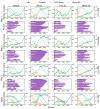
Effects of doubling each of the major energy terms on the lateral pulling of GlpG. The interaction and force plots for each replica are accompanied by a timeline of helical structure below (purple bars). Columns are forcefield conditions and rows are replicas. First column: standard forcefield; second: H-bond energy doubled; third: non-HB potential doubled; fourth: protein–membrane potential doubled. Last row are the average results of 28 replicas.

References

    1. Plaxco K.W., Simons K.T., Baker D. Contact Order, Transition State Placement and the Refolding Rates of Single Domain Proteins. J. Mol. Biol. 1998;277:985–994. doi: 10.1006/jmbi.1998.1645. - DOI - PubMed
    1. Sosnick T.R., Mayne L., Englander S.W. Molecular Collapse: The Rate-Limiting Step in Two-State Cytochrome c Folding. Proteins. 1996;24:413–426. doi: 10.1002/(SICI)1097-0134(199604)24:4<413::AID-PROT1>3.0.CO;2-F. - DOI - PubMed
    1. Pandit A.D., Jha A., Freed K.F., Sosnick T.R. Small Proteins Fold through Transition States with Native-like Topologies. J. Mol. Biol. 2006;361:755–770. doi: 10.1016/j.jmb.2006.06.041. - DOI - PubMed
    1. Baxa M.C., Sosnick T.R. Engineered Metal-Binding Sites to Probe Protein Folding Transition States: Psi Analysis. Methods Mol. Biol. 2022;2376:31–63. doi: 10.1007/978-1-0716-1716-8_2. - DOI - PubMed
    1. Fang X.W., Thiyagarajan P., Sosnick T.R., Pan T. The Rate-Limiting Step in the Folding of a Large Ribozyme without Kinetic Traps. Proc. Natl. Acad. Sci. USA. 2002;99:8518–8523. doi: 10.1073/pnas.142288399. - DOI - PMC - PubMed

Substances

LinkOut - more resources