Review
. 2023 Jan 31;24(3):2697.
doi: 10.3390/ijms24032697.

Application of Antisense Conjugates for the Treatment of Myotonic Dystrophy Type 1

Affiliations
Review

Application of Antisense Conjugates for the Treatment of Myotonic Dystrophy Type 1

Jessica Stoodley et al. Int J Mol Sci. .

Abstract

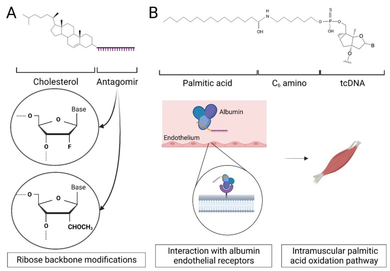
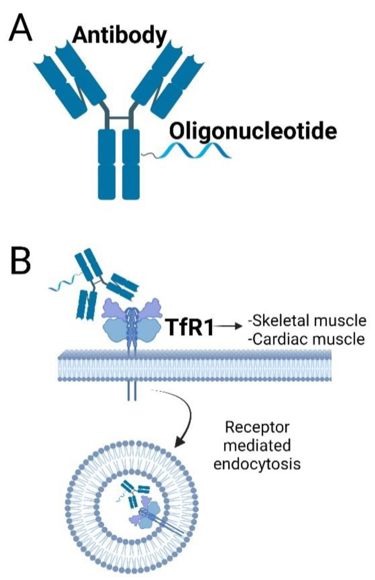
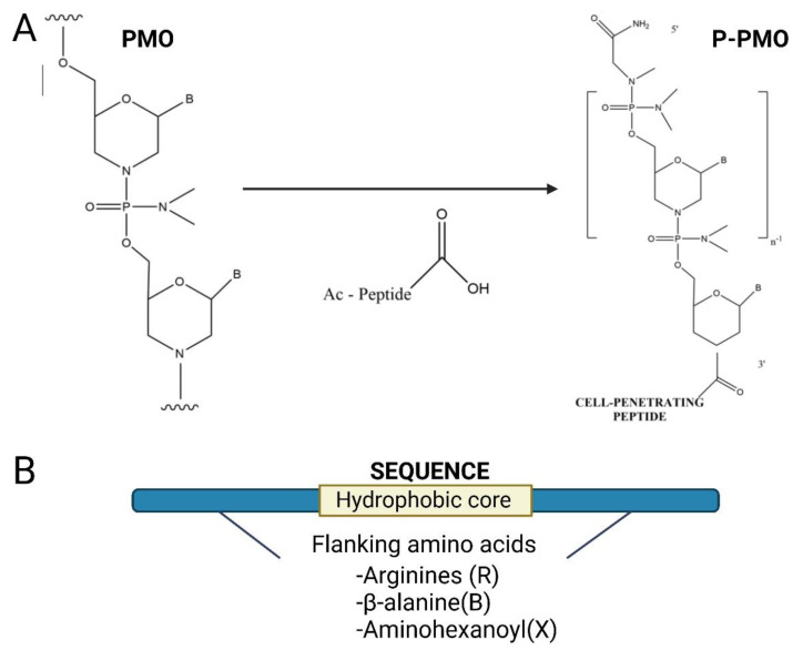
Myotonic dystrophy type 1 (DM1) is one of the most common muscular dystrophies and can be potentially treated with antisense therapy decreasing mutant DMPK, targeting miRNAs or their binding sites or via a blocking mechanism for MBNL1 displacement from the repeats. Unconjugated antisense molecules are able to correct the disease phenotype in mouse models, but they show poor muscle penetration upon systemic delivery in DM1 patients. In order to overcome this challenge, research has focused on the improvement of the therapeutic window and biodistribution of antisense therapy using bioconjugation to lipids, cell penetrating peptides or antibodies. Antisense conjugates are able to induce the long-lasting correction of DM1 pathology at both molecular and functional levels and also efficiently penetrate hard-to-reach tissues such as cardiac muscle. Delivery to the CNS at clinically relevant levels remains challenging and the use of alternative administration routes may be necessary to ameliorate some of the symptoms experienced by DM1 patients. With several antisense therapies currently in clinical trials, the outlook for achieving a clinically approved treatment for patients has never looked more promising.

Keywords: antibody; bridged nucleic acids; cell-penetrating peptide; lipids; muscle; myotonic dystrophy; oligonucleotides; splicing.


Conflict of interest statement

MJAW and MAV are co-inventors of patents licensed to PepGen, a company dedicated to the peptide-based enhancement of delivery of therapeutic oligonucleotide analogs.

Figures

Figure 1
Figure 1
Antisense molecules can be conjugated to lipids to improve their pharmacokinetic properties. (A) Single-stranded RNA analogues complementary to miRNAs (AntagomiR) conjugated to cholesterol are delivered to non-CNS tissues at clinically relevant concentrations. Crossing the BBB into the CNS can be improved by conjugating the lipid to a DNA/RNA heteroduplex oligonucleotide [71]. (B) Conjugation of tricyclo-DNA (tcDNA) [72] ASOs with fatty acids such as palmitic acid has also been shown to improve ASO distribution by binding to serum albumin and indirectly with endothelial surface receptors facilitating transport across the continuous capillary endothelium in muscle.
Figure 2
Figure 2
Antisense Oligonucleotides can be conjugated to antibodies targeting the Transferrin Receptor 1 (TfR1) and penetrate cells by receptor-mediated endocytosis. (A) Schematic design of an antibody–ASO conjugate. The ASO is conjugated directly to the antibody using covalent unions. (B) Antibodies targeting TfR1, a highly expressed cell-surface receptor of DM1 in target tissues such as skeletal and cardiac muscles, have been used to deliver antisense cargo. The AOC enters the cell by receptor-mediated endocytosis resulting on an enhanced ASO delivery. The affinity of TfR1antibodies can be fine-tuned to maximise the interaction with receptors in the BBB allowing for the compound to be delivered to the CNS (in mice) after systemic administration [85].
Figure 3
Figure 3
Antisense oligonucleotides can be conjugated to cell-penetrating peptides (CPPs) to increase cellular uptake. (A) CPPs can be directly conjugated to charge-neutral oligonucleotides, such as PMOs or PNAs using direct covalent unions [35]. (B) The peptide sequence is usually positively charged and rich in arginine. Here, a cell-penetrating peptide comprising a hydrophobic core and two flanking regions enriched with cationic amino acids such as arginine or B-alanine is shown.

References

    1. Norwood F.L.M., Harling C., Chinnery P.F., Eagle M., Bushby K., Straub V. Prevalence of genetic muscle disease in Northern England: In-depth analysis of a muscle clinic population. Brain. 2009;132:3175–3186. doi: 10.1093/brain/awp236. - DOI - PMC - PubMed
    1. Siciliano G., Manca M., Gennarelli M., Rocchi A., Iudice A., Miorin M., Mostacciuolo M. Epidemiology of myotonic dystrophy in Italy: Re-apprisal after genetic diagnosis. Clin. Genet. 2001;59:344–349. doi: 10.1034/j.1399-0004.2001.590508.x. - DOI - PubMed
    1. Johnson N.E., Butterfield R.J., Mayne K., Newcomb T., Imburgia C., Dunn D., Duval B., Feldkamp M.L., Weiss R.B. Population-Based Prevalence of Myotonic Dystrophy Type 1 Using Genetic Analysis of Statewide Blood Screening Program. Neurology. 2021;96:e1045–e1053. doi: 10.1212/WNL.0000000000011425. - DOI - PMC - PubMed
    1. Echenne B., Bassez G. Congenital and infantile myotonic dystrophy. Handb. Clin. Neurol. 2013;113:1387–1393. doi: 10.1016/B978-0-444-59565-2.00009-5. - DOI - PubMed
    1. Mahadevan M., Tsilfidis C., Sabourin L., Shutler G., Amemiya C., Jansen G., Neville C., Narang M., Barceló J., O’Hoy K. Myotonic dystrophy mutation: An unstable CTG repeat in the 3′ untranslated region of the gene. Science. 1992;255:1253–1255. doi: 10.1126/science.1546325. - DOI - PubMed

Substances