. 2023 Jan 31;24(3):2711.
doi: 10.3390/ijms24032711.

Moderating Gut Microbiome/Mitochondrial Axis in Oxazolone Induced Ulcerative Colitis: The Evolving Role of β-Glucan and/or, Aldose Reductase Inhibitor, Fidarestat

Affiliations

Moderating Gut Microbiome/Mitochondrial Axis in Oxazolone Induced Ulcerative Colitis: The Evolving Role of β-Glucan and/or, Aldose Reductase Inhibitor, Fidarestat

Omnia Safwat El-Deeb et al. Int J Mol Sci. .

Abstract

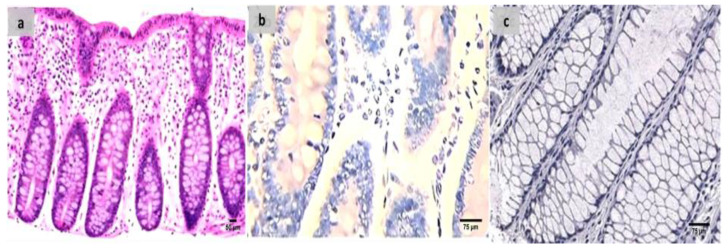
A mechanistic understanding of the dynamic interactions between the mitochondria and the gut microbiome is thought to offer innovative explanations for many diseases and thus provide innovative management approaches, especially in GIT-related autoimmune diseases, such as ulcerative colitis (UC). β-Glucans, important components of many nutritious diets, including oats and mushrooms, have been shown to exhibit a variety of biological anti-inflammatory and immune-modulating actions. Our research study sought to provide insight into the function of β-glucan and/or fidarestat in modifying the microbiome/mitochondrial gut axis in the treatment of UC. A total of 50 Wistar albino male rats were grouped into five groups: control, UC, β-Glucan, Fidarestat, and combined treatment groups. All the groups were tested for the presence of free fatty acid receptors 2 and 3 (FFAR-2 and -3) and mitochondrial transcription factor A (TFAM) mRNA gene expressions. The reactive oxygen species (ROS), mitochondrial membrane potential (MMP), and ATP content were found. The trimethylamine N-oxide (TMAO) and short-chain fatty acid (SCFA) levels were also examined. Nuclear factor kappa β (NF-kβ), nuclear factor (erythroid-2)-related factor 2 (Nrf2) DNA binding activity, and peroxisome proliferator-activated receptor gamma co-activator-1 (PGC-1) were identified using the ELISA method. We observed a substantial increase FFAR-2, -3, and TFAM mRNA expression after the therapy. Similar increases were seen in the ATP levels, MMP, SCFA, PGC-1, and Nrf2 DNA binding activity. The levels of ROS, TMAO, and NF-kβ, on the other hand, significantly decreased. Using β-glucan and fidarestat together had unique therapeutic benefits in treating UC by focusing on the microbiota/mitochondrial axis, opening up a new avenue for a potential treatment for such a complex, multidimensional illness.

Keywords: SCFA; fidarestat; microbiota; mitochondria; ulcerative colitis; ꞵ-glucan.


Conflict of interest statement

The authors declare no conflict of interest.

Figures

Figure 1
Figure 1
Effect of Oxazolone, β-glucan and fidarestat on relative mRNA expressions of colonic FFAR2, colonic FFAR3, and mitochondrial TFAM. (A). Relative mRNA expression of colonic free fatty acid receptor 2 (FFAR-2), (B). Relative mRNA expression of colonic free fatty acid receptor3 (FFAR3), (C). Relative mRNA expression of mitochondrial transcription factor A (TFAM). Data are expressed as mean ± SD. a p < 0.05 vs. control group, b p < 0.05 vs. UC group, c p < 0.05 vs. β-glucan-treated group and d p < 0.05 vs. Fidarestat-treated group.
Figure 2
Figure 2
(a) Normal control colon specimen (H&E ×100), (b) normal colon stained by calprotectin showed negative expression (×200), (c): normal colon stained by S100A12 showed negative expression (×200).
Figure 3
Figure 3
(a) A case of UC Geboes score 4.4 showed marked epithelial injury, cryptitis and ulcerations (H&E ×200), (b) the same case showed positive calprotectin expression in the mucosal cells and the stroma (×200), (c) also showed positive S100A12 expression in the mucosal cells (×200).
Figure 4
Figure 4
(a) β-glucan-treated group showing decreased activity into Geboes score 3.1 in the form of cryptitis and neutrophils seen in some of the epithelium (H&E ×200). (b) The same case showed less number of positive mucosal cells for calprotectin expression (×200), (c) also showed weak positive expression of S100A12 (×200).
Figure 5
Figure 5
(a) Fidarestat treated group showing decreased activity into Geboes score of 3.2 in the form of cryptitis and neutrophils seen in most of the epithelium (H&E ×200). (b) The same case showed weak calprotectin but focal expression (×200), (c) also showed weak and focal S100A12 expression (×200).
Figure 6
Figure 6
(a) Combined treated group showing Geboes score of 1.2 in the form of a marked increase in the basal plasma cells only with no crypt abscesses (H&E ×200). (b) The same case showed negative calprotectin expression (×200), (c) also showed negative S100A12 expression (×200).

References

    1. Mottawea W., Chiang C.K., Mühlbauer M., Starr A.E., Butcher J., Abujamel T., Deeke S.A., Brandel A., Zhou H., Shokralla S., et al. Altered intestinal microbiota–host mitochondria crosstalk in new onset Crohn’s disease. Nat. Commun. 2016;7:13419. doi: 10.1038/ncomms13419. - DOI - PMC - PubMed
    1. Saint-Georges-Chaumet Y., Edeas M. Microbiota–mitochondria inter-talk: Consequence for microbiota–host interaction. Pathog. Dis. 2016;74:ftv096. doi: 10.1093/femspd/ftv096. - DOI - PubMed
    1. Clark A., Mach N. The crosstalk between the gut microbiota and mitochondria during exercise. Front. Physiol. 2017;8:319. doi: 10.3389/fphys.2017.00319. - DOI - PMC - PubMed
    1. Circu M.L., Aw T.Y. Seminars in Cell & Developmental Biology. Volume 23. Academic Press; Cambridge, MA, USA: 2012. Intestinal redox biology and oxidative stress; pp. 729–737. - PMC - PubMed
    1. Bär F., Bochmann W., Widok A., Von Medem K., Pagel R., Hirose M., Yu X., Kalies K., König P., Böhm R., et al. Mitochondrial gene polymorphisms that protect mice from colitis. Gastroenterology. 2013;145:1055–1063. doi: 10.1053/j.gastro.2013.07.015. - DOI - PubMed