. 2023 Feb 1;24(3):2732.
doi: 10.3390/ijms24032732.

Dysregulation of Gene Expression of Key Signaling Mediators in PBMCs from People with Type 2 Diabetes Mellitus

Affiliations

Dysregulation of Gene Expression of Key Signaling Mediators in PBMCs from People with Type 2 Diabetes Mellitus

Nilofer Qureshi et al. Int J Mol Sci. .

Abstract

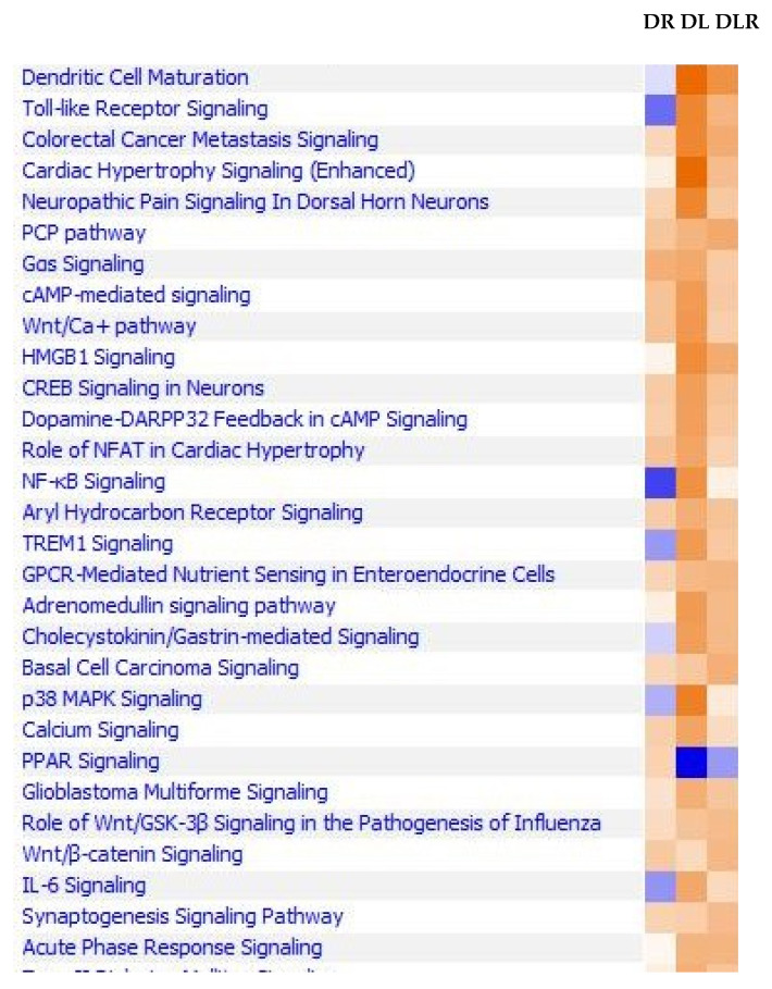
Diabetes is currently the fifth leading cause of death by disease in the USA. The underlying mechanisms for type 2 Diabetes Mellitus (DM2) and the enhanced susceptibility of such patients to inflammatory disorders and infections remain to be fully defined. We have recently shown that peripheral blood mononuclear cells (PBMCs) from non-diabetic people upregulate expression of inflammatory genes in response to proteasome modulators, such as bacterial lipopolysaccharide (LPS) and soybean lectin (LEC); in contrast, resveratrol (RES) downregulates this response. We hypothesized that LPS and LEC will also elicit a similar upregulation of gene expression of key signaling mediators in (PBMCs) from people with type 2 diabetes (PwD2, with chronic inflammation) ex vivo. Unexpectedly, using next generation sequencing (NGS), we show for the first time, that PBMCs from PwD2 failed to elicit a robust LPS- and LEC-induced gene expression of proteasome subunit LMP7 (PSMB8) and mediators of T cell signaling that were observed in non-diabetic controls. These repressed genes included: PSMB8, PSMB9, interferon-γ, interferon-λ, signal-transducer-and-activator-of-transcription-1 (STAT1), human leukocyte antigen (HLA DQB1, HLA DQA1) molecules, interleukin 12A, tumor necrosis factor-α, transporter associated with antigen processing 1 (TAP1), and several others, which showed a markedly weak upregulation with toxins in PBMCs from PwD2, as compared to those from non-diabetics. Resveratrol (proteasome inhibitor) further downregulated the gene expression of these inflammatory mediators in PBMCs from PwD2. These results might explain why PwD2 may be susceptible to infectious disease. LPS and toxins may be leading to inflammation, insulin resistance, and thus, metabolic changes in the host cells.

Keywords: IFN-γ; LPS; NGS; NO; cytokines; lectins; resveratrol; signal transduction; type 2 diabetes.


Conflict of interest statement

The authors declare no conflict of interest.

Figures

Figure 1
Figure 1
Cell functions modulated by RES, LPS, and LPS + RES in PBMCs from controls and PwD2. Human PBMCs from non-diabetic and PwD2 were treated with three different treatments and the vehicle control for 3 h. The RNA samples from controls and PwD2 were extracted from the cells and analyzed using RNAseq. These data were first extracted using the DEG analysis, followed by ingenuity pathways analysis. Cell functions for RES (R), LPS (L), LPS + RES (L + R), DRES (DR), DLPS (DL), and DLPS + RES (DL + R). Comparisons were made using their respective controls. Dark orange color denotes maximal effect, while dark blue color denotes minimal effect.
Figure 2
Figure 2
The role of RES, LPS, LPS + RES, LEC10, LEC50, on gene expression of mediators involved in signaling pathways in PBMCs from non-diabetic controls and PwD2. Human PBMCs from controls and PwD2 were treated and compared with the vehicle control for 3 h. RNA was extracted from the cells and analyzed using RNAseq. These data were first extracted using the DEG analysis, followed by ingenuity pathways analysis. Z scores are plotted against the signaling pathways. Nondiabetics RES (R); LPS (L); LPS + RES (L + R); LEC10; LEC50 (blue colored bars; Diabetics, RES (DR); LPS (DL); LPS + RES (DL + R); DLEC10 (LEC10); and DLEC50 (LEC50). (AF) Signaling pathways modulated by LPS, LEC, RES in PBMCs from non-diabetic control (blue bars) vs. PwD2 (red colored bars).
Figure 3
Figure 3
The mediators involved in signaling pathways that were downregulated by RES. The role of RES (DR), LPS (DL), and LPS + RES (DLR) on gene expression of mediators involved in the agonist-induced signal transduction in treated PBMC from PwD2 are described in the legend to Figure 2. RNAseq data were first extracted using the DEG analysis and analyzed by ingenuity pathways analysis. Z scores were plotted against the signaling pathways, where dark orange denotes maximum activation and dark blue maximum repression.
Figure 4
Figure 4
(AD) Modulation of proteasome subunits: the effect of LPS, LEC10, LEC50, and RES on gene expression of proteasome’s subunits X, Y, Z, LMP7, LMP2, and LMP10 was determined in PBMCs from PwD2 and non-diabetic controls. Gene expression of subunits X and LMP10 was not induced in human PBMCs from non-diabetic controls and PwD2. Human PBMCs were treated with compounds and vehicle for 3h. RNA was extracted from the cells and analyzed using NGS. These data were extracted using the DEG analysis. Z score values were plotted against the proteasome subunits. Control PBMCs (C), RES (R); LPS (L); LPS + RES (L + R); LEC10; LEC50; and Diabetic PBMCs: RES (DR); LPS (DL); LPS + RES (DL + R); LEC10 (DLEC10); and LEC50 (DLEC50). Blue bars indicate values for gene expression of non-diabetic controls [54] and red bars indicate values for PwD2.
Figure 5
Figure 5
IFN-induced genes were downregulated in PBMCs from PwD2 as compared to non-diabetic controls in response to LPS, except for IFN-β. RES showed an inhibition or no change in gene expression, whereas LPS and LEC10 showed an upregulation of these genes. CRES, CLPS, and CLEC50 (controls) and DRES, DLPS, and DLEC50 (PwD2) were compared. (A) Non-diabetic control and (B) PwD2.
Figure 6
Figure 6
Cytokine and other inflammation-linked genes modulated by RES (R), LPS (L), LPS + RES (LPS + R, LR), LEC10 (lectin 10 μg/mL) and LEC50 (lectin 50 μg/mL) in PBMCs from non-diabetic controls (blue bars); and DRES (DR), DLPS (DL), DLPS + RES (DL + R), DLEC10 (lectin 10 μg/mL) and DLEC50 (lectin 50 μg/mL) in PBMCs from PwD2 (red bars). Cytokine genes are differentially regulated in PBMCs of non-diabetic controls and PwD2. (A). IFN-γ, (B). IL-20, (C). IL-12A, (D). INSR, (E). INSM1, (F). INSRR, (G). IL-2, (H). TLR9, and (I). IFN-λ.
Figure 7
Figure 7
RNA was extracted from cells and gene expression was analyzed using qRT-PCR for validation of NGS data (Expt. 5). (A,B) TNF-α and iNOS genes modulated by medium control, LEC10 and LEC50, (C,D) IL-4 and IFN-γ genes modulated by different treatments in PBMCs of non-diabetics and PwD2.
Figure 8
Figure 8
PBMCs were treated with either vehicle, RES, LPS, LPS + RES, or LEC10. RNA was extracted from cells and gene expression of IFN-γ and TNF-α was analyzed using qRT-PCR. Mean ± SEM. In these graphs, C, non-diabetic control was used to calculate 2−ΔΔCT for non-diabetics, and D control was used for diabetics. LPS did not induce gene expression of IFN-γ as robustly in PBMCs from PwD2, compared to non-diabetic controls. Values in a column sharing a common asterisk with proteasome modulators were significantly different at *, **, *** p < 0.024, 0.007, 0.0001, using 1-way Anova. 2−ΔΔCT = Relative quantification RQ. (Experiments 1–4 are described in the Section 4).
Figure 9
Figure 9
The interferon signaling pathway in human PBMC. The interferon signaling pathway plays a critical role in the induction of immune response by LPS or viruses: the STAT1 and STAT2 transcription factors play a major role in the transcription of interferon-activated genes. (A). LPS-non-diabetic and (B). LPS-diabetic. Red color denotes activation of genes, green color denotes repression of genes, and grey color denotes no change in gene expression.
Figure 10
Figure 10
A simplistic model for LPS-induced genes in non-diabetic vs. PwD2. Figure shows the modulation of gene expression in treated PBMCs of PwD2/non-diabetics with RES, LPS, and LEC. Respective controls were used for analyses. A simplistic model for activation of innate and acquired immunity in PBMCs based on LPS-mediated gene expression. The role of RES, LPS, LPS + RES, and LEC10 on the innate and acquired immunity in human PBMCs from non-diabetic controls and PwD2 was determined.

Similar articles

References

    1. Singh A.K., Gupta R., Ghosh A., Misra A. Diabetes in COVID-19: Prevalence, pathophysiology, prognosis and practical considerations. Diabetes Metab. Syndr. Clin. Res. Rev. 2020;14:303–310. - PMC - PubMed
    1. Jesenak M., Brndiarova M., Urbancikova I., Rennerova Z., Vojtkova J., Bobcakova A., Ostro R., Banovcin P. Immune parameters, and COVID-19 infection-associations with clinical severity and diseases prognosis. Front. Cell. Infect. Microbiology. 2020;10:364. doi: 10.3389/fcimb.2020.00364. - DOI - PMC - PubMed
    1. Krug E.G. Trends in diabetes: Sounding the alarm. Lancet. 2016;387:1485–1486. doi: 10.1016/S0140-6736(16)30163-5. - DOI - PubMed
    1. Wing S.S. The UPS in diabetes and obesity. BMC Biochem. 2008;9:S6. doi: 10.1186/1471-2091-9-S1-S6. - DOI - PMC - PubMed
    1. Siddiqui A.A., Siddiqui S.A., Ahmad S., Siddiqui S., Ahsan I., Sahu K. Diabetes: Mechanism, pathophysiology, and management-A review. Int. J. Drug Dev. Res. 2013;5:1–23.

MeSH terms