Review
. 2023 Feb 1;24(3):2825.
doi: 10.3390/ijms24032825.

Localisation of Intracellular Signals and Responses during Phagocytosis

Affiliations
Review

Localisation of Intracellular Signals and Responses during Phagocytosis

Maurice B Hallett. Int J Mol Sci. .

Abstract

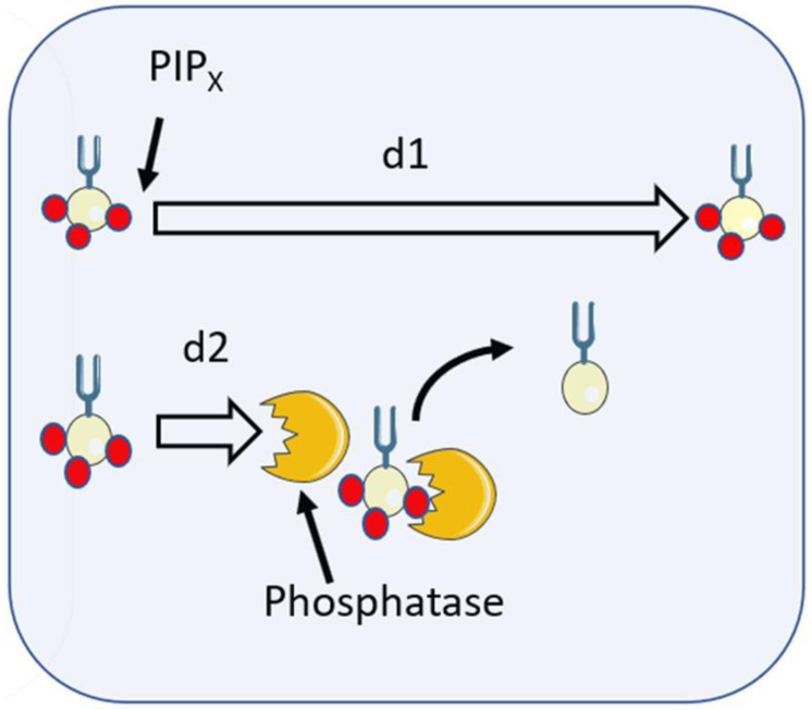
Phagocytosis is one of the most polarised of all cellular activities. Both the stimulus (the target for phagocytosis) and the response (its internalisation) are focussed at just one part of the cell. At the locus, and this locus alone, pseudopodia form a phagocytic cup around the particle, the cytoskeleton is rearranged, the plasma membrane is reorganised, and a new internal organelle, the phagosome, is formed. The effect of signals from the stimulus must, thus, both be complex and yet be restricted in space and time to enable an effective focussed response. While many aspects of phagocytosis are being uncovered, the mechanism for the restriction of signalling or the effects of signalling remains obscure. In this review, the details of the problem of restricting chemical intracellular signalling are presented, with a focus on diffusion into the cytosol and of signalling lipids along the plasma membrane. The possible ways in which simple diffusion is overcome so that the restriction of signalling and effective phagocytosis can be achieved are discussed in the light of recent advances in imaging, biophysics, and cell biochemistry which together are providing new insights into this area.

Keywords: Ca2+; cell signalling; cytoskeleton; neutrophils; phagocytosis; phospholipids.


Conflict of interest statement

The author declares no conflict of interest.

Figures

Figure 1
Figure 1
The effect of diffusion of the spread of chemical signals. (a) shows the 1-dimenional distribution of the concentration of molecules (or ions such as Ca2+) from a point source (at zero distance) with time using the equation shown, where C=1-dimensional concentration (e.g., molecules or ions/μm); n=the number of molecules initially at the source (i.e., at time zero and distance zero): x= distance from the initial source (μm); and D=diffusion constant for the molecule (taken here as 1 μm2/s). The concentration of the molecules or ions in the profiles is shown on the vertical axis in arbitrary units. Each curve shows the concentration profile at the time indicated. (b) The concentration profile of the signalling molecules at a single time point is shown, with the “diffusion length” (=2 √(Dt) shown for comparison. (c) The effectiveness of the signalling-molecule concentration depends on the target requirement, whether it is activated by a low concentration (shown as 1), medium concentration (shown as 2), or high concentration (shown as 3). The “zone of signalling” is reduced by an increased requirement of the target for the signalling molecule as shown by the 1-dimensional spread of activated target molecules (dotted lines) and a 2-dimensional “target” representation, where the darker shading indicates more signalling effectiveness.
Figure 2
Figure 2
The Effect of Ca2+ buffering on diffusion length (signalling reach).
Figure 3
Figure 3
The effect of the two routes of Ca2+ signalling on retaining information as to its locus of origin.
Figure 4
Figure 4
The lateral diffusion of signalling lipids at the site of phagocytosis.
Figure 5
Figure 5
PIPx interactions with inositol phosphatases limit diffusion length.

Similar articles

Cited by

References

    1. Uribe-Querol E., Rosales C. Phagocytosis: Our Current Understanding of a Universal Biological Process. Front. Immunol. 2020;11:1066. doi: 10.3389/fimmu.2020.01066. - DOI - PMC - PubMed
    1. Aderem A., Underhill D.M. Mechanisms of Phagocytosis in Macrophages. Ann. Rev. Immunol. 1999;17:593–623. doi: 10.1146/annurev.immunol.17.1.593. - DOI - PubMed
    1. Lee H.-J., Woo Y., Hahn T.-W., Jung Y.M., Jung Y.-J. Formation and Maturation of the Phagosome: A Key Mechanism in Innate Immunity against Intracellular Bacterial Infection. Microorganisms. 2020;8:1298. doi: 10.3390/microorganisms8091298. - DOI - PMC - PubMed
    1. Champion J.A., Mitragotry S. Role of target geometry in phagocytosis. Proc. Natl. Acad. Sci. USA. 2006;103:4930–4934. doi: 10.1073/pnas.0600997103. - DOI - PMC - PubMed
    1. Richards D.M. Receptor Models of Phagocytosis: The Effect of Target Shape. Adv. Expt. Med. Biol. 2020;1246:55–70. - PubMed

LinkOut - more resources