

Brain N-Glycosylation and Lipidomic Profile Changes Induced by a High-Fat Diet in Dyslipidemic Hamsters
- PMID: 36769208
- PMCID: PMC9918045
- DOI: 10.3390/ijms24032883
Brain N-Glycosylation and Lipidomic Profile Changes Induced by a High-Fat Diet in Dyslipidemic Hamsters
Abstract
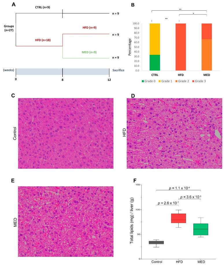
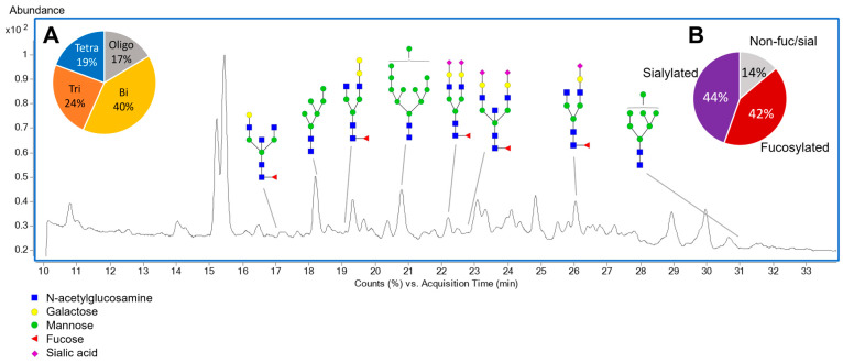
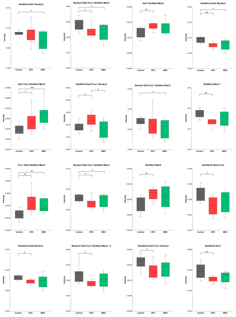
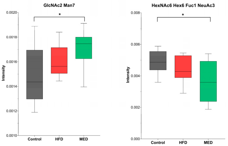
The consumption of diets rich in saturated fats is known to be associated with higher mortality. The adoption of healthy habits, for instance adhering to a Mediterranean diet, has proved to exert a preventive effect towards cardiovascular diseases and dyslipidemia. Little is known about how a suboptimal diet can affect brain function, structure, and the mechanisms involved. The aims of this study were to examine how a high-fat diet can alter the brain N-glycan and lipid profile in male Golden Syrian hamsters and to evaluate the potential of a Mediterranean-like diet to reverse this situation. During twelve weeks, hamsters were fed a normal fat diet (CTRL group), a high-fat diet (HFD group), and a high-fat diet followed by a Mediterranean-like diet (MED group). Out of seventy-two identified N-glycans, fourteen were significant (p < 0.05) between HFD and CTRL groups, nine between MED and CTRL groups, and one between MED and HFD groups. Moreover, forty-nine lipids were altered between HFD and CTRL groups, seven between MED and CTRL groups, and five between MED and HFD groups. Our results suggest that brain N-glycan composition in high-fat diet-fed hamsters can produce events comparable to those found in some neurodegenerative diseases, and may promote brain ageing.
Keywords: N-glycan; brain glycosylation; dyslipidemia; high-fat diet; lipidomics; mass spectrometry; mediterranean diet.
Conflict of interest statement
The authors declare no conflict of interest.
Figures
References
-
- Afshin A., Sur P.J., Fay K.A., Cornaby L., Ferrara G., Salama J.S., Mullany E.C., Abate K.H., Abbafati C., Abebe Z., et al. Health effects of dietary risks in 195 countries, 1990–2017: A systematic analysis for the Global Burden of Disease Study 2017. Lancet. 2019;393:1958–1972. doi: 10.1016/S0140-6736(19)30041-8. - DOI - PMC - PubMed
-
- Karlsson H.K., Tuominen L., Tuulari J.J., Hirvonen J., Parkkola R., Helin S., Salminen P., Nuutila P., Nummenmaa L. Obesity is associated with decreased µ-opioid but unaltered dopamine D2 receptor availability in the brain. J. Neurosci. 2015;35:3959–3965. doi: 10.1523/JNEUROSCI.4744-14.2015. - DOI - PMC - PubMed
MeSH terms
Substances
Grants and funding
LinkOut - more resources
Full Text Sources
