. 2023 Feb 2;24(3):2922.
doi: 10.3390/ijms24032922.

Protein Susceptibility to Peroxidation by 4-Hydroxynonenal in Hereditary Hemochromatosis

Affiliations

Protein Susceptibility to Peroxidation by 4-Hydroxynonenal in Hereditary Hemochromatosis

Sandra Sánchez-Jaut et al. Int J Mol Sci. .

Abstract

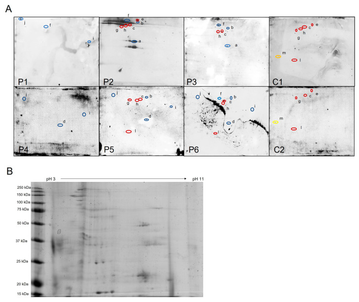
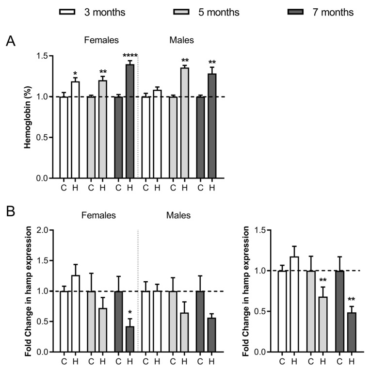
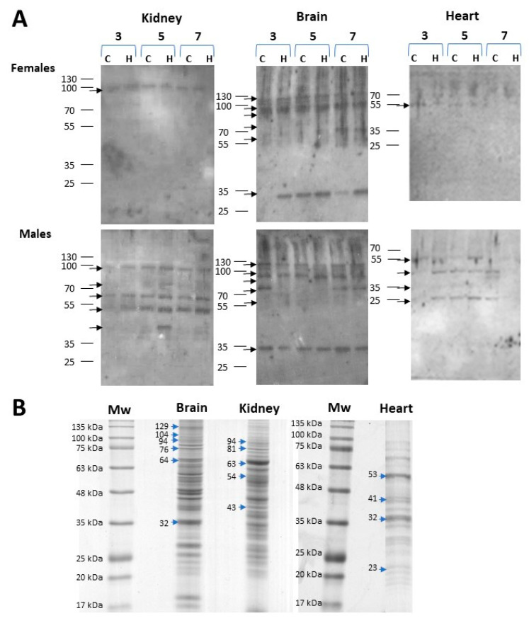
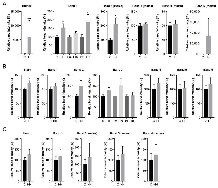
Iron overload caused by hereditary hemochromatosis (HH) increases free reactive oxygen species that, in turn, induce lipid peroxidation. Its 4-hydroxynonenal (HNE) by-product is a well-established marker of lipid peroxidation since it reacts with accessible proteins with deleterious consequences. Indeed, elevated levels of HNE are often detected in a wide variety of human diseases related to oxidative stress. Here, we evaluated HNE-modified proteins in the membrane of erythrocytes from HH patients and in organs of Hfe-/- male and female mice, a mouse model of HH. For this purpose, we used one- and two-dimensional gel electrophoresis, immunoblotting and MALDI-TOF/TOF analysis. We identified cytoskeletal membrane proteins and membrane receptors of erythrocytes bound to HNE exclusively in HH patients. Furthermore, kidney and brain of Hfe-/- mice contained more HNE-adducted protein than healthy controls. Our results identified main HNE-modified proteins suggesting that HH favours preferred protein targets for oxidation by HNE.

Keywords: 4-hydroxynonenal (HNE); Hfe−/− mouse; erythrocyte membrane proteins; hemochromatosis; lipid peroxidation; oxidative stress; protein modification.


Conflict of interest statement

The authors declare no conflict of interest.

Figures

Figure 1
Figure 1
Identification of HNE-modified proteins from erythrocyte membrane of HH patients. (A) Western blots of HH patients (P1–P6) and healthy controls (C1 and C2). Protein spots identified by MALDI-TOF-TOF are designated with “a–m” letters and encircled with colours: red are spots present in patients and controls; blue spots are present only in HH patients; and yellow spots are only in controls. (B) Representative two-dimensional Coomassie brilliant blue-stained gel with total erythrocyte membrane proteins (Control sample 1). 4-hydroxynonenal (HNE), Hereditary hemochromatosis (HH).
Figure 2
Figure 2
Haemoglobin levels and mRNA hepcidin expression in Hfe−/− mice. (A) Normalised haemoglobin concentration in peripheral blood. (B) mRNA expression of hepatic hepcidin at 3, 5 and 7 months in Hfe−/− (H) and wild type (C) female and male mice. Hamp (hepcidin gene) mRNA expression is also shown without gender separation (bottom right). The data for each Hfe−/− mouse were normalised to controls of the same age and sex (indicated by dotted lines). Representative graphs show mean ± SEM from 2 independent experiments (each experiment n = 6 per bar/sex group). Statistical significance is indicated as **** p < 0.0001, ** p < 0.01 and * p < 0.05 in comparison to age- and sex-matched controls.
Figure 3
Figure 3
HNE-modified proteins in organs from female and male Hfe−/− mice. Homogenates from each organ were separately resolved on 12% polyacrylamide gels. HNE (4-hydroxynonenal) modified proteins were detected by Western blot using HNE specific antibodies. (A) Western blot analysis of HNE adducts in kidney, brain, and heart proteins from female and male control (C) and Hfe−/− (H) mice at 3, 5 and 7 months of age. Images are representative of n = 3 per group. (B) Coomassie brilliant blue-stained gels where HNE-immunoreactive bands protein bands were excised. Arrows indicate the protein bands that were excised from the gel and the molecular weight are expressed in KDa on the left side of the arrow.
Figure 4
Figure 4
Densitometric analyses of 4-hydroxynonenal (HNE)-protein signals. Global and individual intensities of bands as shown in Figure 3A and identified in Table 2 from (A) kidney, (B) brain and (C) heart of control (C) and Hfe−/− (H) mice. Analysis of male (m) and female (f) mice is shown separately only in cases of statistically significant results. When band analysis is shown for males only, it is indicated in brackets (males) at the top. Total Hfe−/− mice or control n = 18; male/female Hfe−/− or control mice n = 9. Values are expressed as relative band intensity (%) considering 100% the mean value of n = 18 control mice or n = 9 male/female control mice as indicated (mean ± SEM). Statistical comparisons were performed between groups using Student unpaired t-tests or Mann–Whitney U-tests. * p < 0.05, *** p ≤ 0.001.
Figure 5
Figure 5
Comparative liver mRNA expression of tnf, il6, gpx1 and sod2. Relative expression of tumour necrosis factor-α (tnf), interleukin 6 (il6), glutathione peroxidase-1 (gpx1) and superoxide dismutase 2 (sod2) in female and male 3-, 5- and 7-month-old Hfe−/− mice (H) and healthy controls (C) was calculated by the 2−ΔΔCT method. The results are expressed as arbitrary units normalised to β-actin to correct for mRNA quantity and integrity. The dotted lines represent normalized values of healthy controls. Data are shown as mean ± SEM, each with n = 6 per group, where * p ≤ 0.05 comparing to same-age controls, and ## p < 0.01 compared to same age Hfe−/− mice but different gender.

References

    1. Papanikolaou G., Samuels M.E., Ludwig E.H., MacDonald M.L.E., Franchini P.L., Dubé M.-P., Andres L., MacFarlane J., Sakellaropoulos N., Politou M., et al. Mutations in HFE2 cause iron overload in chromosome 1q–linked juvenile hemochromatosis. Nat. Genet. 2004;36:77–82. doi: 10.1038/ng1274. - DOI - PubMed
    1. Roetto A., Papanikolaou G., Politou M., Alberti F., Girelli D., Christakis J., Loukopoulos D., Camaschella C. Mutant antimicrobial peptide hepcidin is associated with severe juvenile hemochromatosis. Nat. Genet. 2003;33:21–22. doi: 10.1038/ng1053. - DOI - PubMed
    1. Roetto A., Totaro A., Piperno A., Piga A., Longo F., Garozzo G., Calì A., De Gobbi M., Gasparini P., Camaschella C. New mutations inactivating transferrin receptor 2 in hemochromatosis type. Blood. 2001;97:2555–2560. doi: 10.1182/blood.V97.9.2555. - DOI - PubMed
    1. Njajou O.T., Vaessen N., Joosse M., Berghuis B., Van Dongen J.W.F., Breuning M.H., Snijders P.J.L.M., Rutten W.P.F., Sandkuijl L.A., Oostra B.A., et al. A mutation in SLC11A3 is associated with autosomal dominant hemochromatosis. Nat. Genet. 2001;28:213–214. doi: 10.1038/90038. - DOI - PubMed
    1. Ayala A., Muñoz M.F., Argüelles S. Lipid peroxidation: Production, metabolism, and signaling mechanisms of malondialdehyde and 4-hydroxy-2-nonenal. Oxid. Med. Cell. Longev. 2014;2014:360438. doi: 10.1155/2014/360438. - DOI - PMC - PubMed

LinkOut - more resources