. 2023 Feb 3;24(3):3035.
doi: 10.3390/ijms24033035.

Supt16 Haploinsufficiency Impairs PI3K/AKT/mTOR/Autophagy Pathway in Human Pluripotent Stem Cells Derived Neural Stem Cells

Affiliations

Supt16 Haploinsufficiency Impairs PI3K/AKT/mTOR/Autophagy Pathway in Human Pluripotent Stem Cells Derived Neural Stem Cells

Junwen Wang et al. Int J Mol Sci. .

Abstract

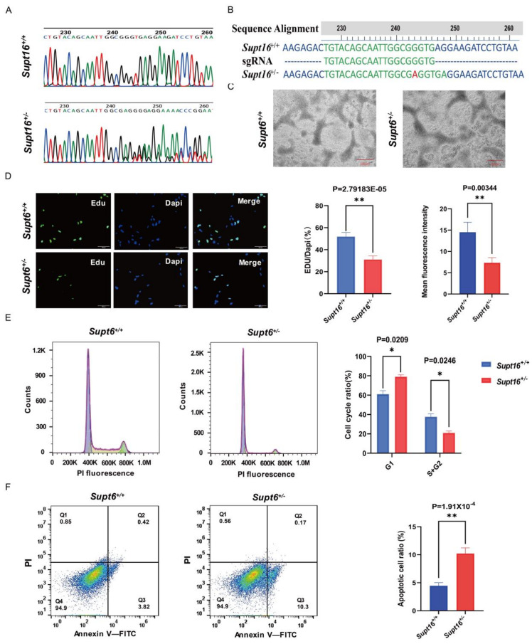
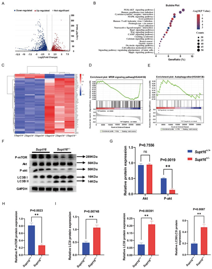
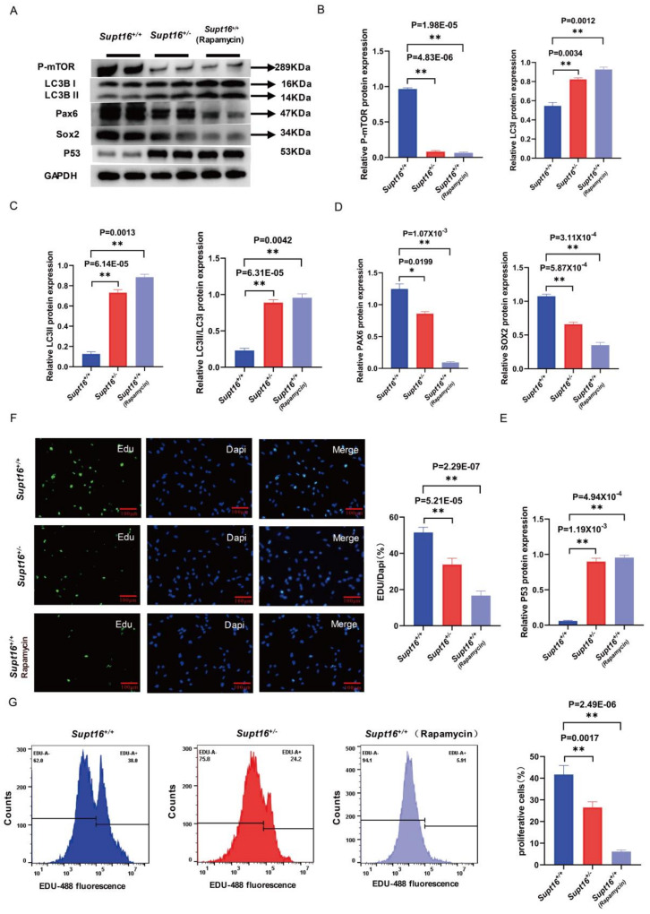
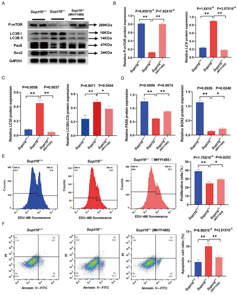
The maintenance of neural stem cells (NSCs) plays a critical role in neurodevelopment and has been implicated in neurodevelopmental disorders (NDDs). However, the underlying mechanisms linking defective human neural stem cell self-renewal to NDDs remain undetermined. Our previous study found that Supt16 haploinsufficiency causes cognitive and social behavior deficits by disrupting the stemness maintenance of NSCs in mice. However, its effects and underlying mechanisms have not been elucidated in human neural stem cells (hNSCs). Here, we generated Supt16+/- induced pluripotent stem cells (iPSCs) and induced them into hNSCs. The results revealed that Supt16 heterozygous hNSCs exhibit impaired proliferation, cell cycle arrest, and increased apoptosis. As the RNA-seq analysis showed, Supt16 haploinsufficiency inhibited the PI3K/AKT/mTOR pathway, leading to rising autophagy, and further resulted in the dysregulated expression of multiple proteins related to cell proliferation and apoptotic process. Furthermore, the suppression of Supt16 heterozygous hNSC self-renewal caused by autophagy activation could be rescued by MHY1485 treatment or reproduced in rapamycin-treated hNSCs. Thus, our results showed that Supt16 was essential for hNSC self-renewal and its haploinsufficiency led to cell cycle arrest, impaired cell proliferation, and increased apoptosis of hNSCs by regulating the PI3K/AKT/mTOR/autophagy pathway. These provided a new insight to understand the causality between the Supt16 heterozygous NSCs and NDDs in humans.

Keywords: Supt16; autophagy; human neural stem cells (hNSCs); neurodevelopmental disorders (NDDs); proliferation.


Conflict of interest statement

The authors declare no conflict of interest.

Figures

Figure 1
Figure 1
Supt16 haploinsufficiency disrupts the self−renewal of human iPSC−derived NSCs. (A) Sanger sequencing of Supt16 heterozygous hNSCs clone after CRISPR/Cas9−mediated. (B) Supt16+/− hNSCs exhibit a point mutation in the form of an adenine nucleotide insertion (red hyphen). (C) Representative pictures of neural rosettes derived from Supt16+/− haploinsufficiency and wild-type iPSCs; scale bar, 100 μm. (D) Representative immunofluorescence images of EdU incorporation assay (n = 3 samples per genotype. Green: EdU−positive cells. Blue: DAPI. Scale bar, 100 μm). (E) Representative flow cytometry analysis picture of hNSCs cell cycle (left). Quantitative analysis of hNSCs cell cycle distribution (right) (n = 3 samples of each genotype). (F) Representative flow cytometry analysis picture and quantification of hNSCs apoptotic rate (n = 3 samples of each genotype). The error bars represented the mean ± SD and the significance level was calculated by Student’s t−test (two−tailed, equal variance) (ns means not statistically significant, * p < 0.05, ** p < 0.01).
Figure 2
Figure 2
Supt16 haploinsufficiency impaired hNSC self−renewal by activating PI3K/AKT/mTOR−mediated autophagy. (A) Volcano plots showed all differential expression genes detected by DESeq2 in Supt16 heterozygous hNSCs. Each point represents an individual gene, of which 1629 genes are upregulated and 1976 genes are downregulated (p−value < 0.05; |log2foldchange| > 1). n = 3. (B) KEGG pathway analysis found that differential expression genes in Supt16+/− hNSCs are enriched in the PI3K/AKT signaling pathway. (C) Heatmap analysis of PI3K/AKT signaling pathway−related differential expression genes enriched in KEGG analysis. (D) GSEA plot of differentially expressed genes for the list of mTOR signaling pathway genes (NES, normalized enrichment score; FDR, false discovery rate. NES = −1.79, FDR = 0.055). (E) GSEA analysis showing the expression pattern of autophagy−related genes in WT and Supt16+/− hNSCs (NES, normalized enrichment score; FDR, false discovery rate. NES = 1.358, FDR = 0.254). (F) Western blot analysis showing AKT, P−akt, P−mTOR, and LC3 expression in hNSCs treated with different treatments (n = 3 samples per genotype). (G) Quantification analysis of AKT and P−akt protein relative to GAPDH. Data are means ± standard deviation of three independent experiments. (H) The P−mTOR expression was calculated relative to GAPDH. Data are means ± standard deviation of three independent experiments. (I) Quantification of LC3 protein relative to GAPDH. Data are means ± standard deviation of three independent experiments. The error bars represented the mean ± SD and the significance level was calculated by Student’s t-test (two-tailed, equal variance) (ns means not statistically significant, ** p < 0.01).
Figure 3
Figure 3
Wild−type hNSCs treated by rapamycin reproduced the phenotype of Supt16 haploinsufficient hNSCs. (A) Western blot analysis showing P−mTOR, LC3, PAX6, SOX2, and P53 protein expression in hNSCs treated with different treatments (n = 3). (B) Quantification analysis of P−mTOR, LC3I protein relative to GAPDH. Data are means ± standard deviation of three independent experiments. (C) Quantification analysis of LC3II and LC3II/LC3I protein relative to GAPDH. Data are means ± standard deviation of three independent experiments. (D) Quantification analysis of PAX6 and SOX2 proteins relative to GAPDH. Data are means ± standard deviation of three independent experiments. (E) Quantification of P53 protein relative to GAPDH. Data are means ± standard deviation of three independent experiments. (F) Immunofluorescence staining showed that the absence of Supt16 in hNSCs suppressed the proliferation. Rapamycin treatment reproduced the inhibitory effect of Supt16 haploinsufficiency on the proliferation of hNSCs. (G) Cell cycle analysis using PI staining in WT, Supt16 heterozygous and rapamycin-treated WT hNSCs (n = 3). The different cell cycle phases were calculated in FlowJo (v10.4.0). The error bars represented the mean ± SD and the significance level was calculated by Student’s t-test (two-tailed, equal variance) (ns means not statistically significant, * p < 0.05, ** p < 0.01).
Figure 4
Figure 4
MHY1485 treatment rescued the phenotypes of Supt16 haploinsufficient hNSCs. (A) Western blot analysis showed the protein expressions of P−mTOR, LC3, PAX6, and SOX2 in hNSCs treated with different treatments. (B) Representative quantification analysis of P−mTOR and LC3I in different hNSCs. (C) Quantification analysis of LC3II and LC3II/LC3I in different hNSCs. Data are means ± standard deviation of three independent experiments. (D) Quantification analysis of PAX6 and SOX2 in different hNSCs. Data are means ± standard deviation of three independent experiments. (E) Cell cycle analysis performed by flow cytometry to calculate the distribution of cell cycle phase (n = 3). (F) Flow cytometric analysis using PI and Annexin−V double staining showed the cell apoptotic rate in hNSCs treated with different treatments (n = 3). The error bars represented the mean ± SD and the significance level was calculated by Student’s t-test (two-tailed, equal variance) (ns means not statistically significant, * p < 0.05, ** p < 0.01).

Similar articles

Cited by

References

    1. Gilissen C., Hehir-Kwa J.Y., Thung D.T., van de Vorst M., van Bon B.W., Willemsen M.H., Kwint M., Janssen I.M., Hoischen A., Schenck A., et al. Genome sequencing identifies major causes of severe intellectual disability. Nature. 2014;511:344–347. doi: 10.1038/nature13394. - DOI - PubMed
    1. Niemi M.E.K., Martin H.C., Rice D.L., Gallone G., Gordon S., Kelemen M., McAloney K., McRae J., Radford E.J., Yu S., et al. Common genetic variants contribute to risk of rare severe neurodevelopmental disorders. Nature. 2018;562:268–271. doi: 10.1038/s41586-018-0566-4. - DOI - PMC - PubMed
    1. Tărlungeanu D.C., Novarino G. Genomics in neurodevelopmental disorders: An avenue to personalized medicine. Exp. Mol. Med. 2018;50:1–7. doi: 10.1038/s12276-018-0129-7. - DOI - PMC - PubMed
    1. Kriegstein A., Alvarez-Buylla A. The glial nature of embryonic and adult neural stem cells. Annu. Rev. Neurosci. 2009;32:149–184. doi: 10.1146/annurev.neuro.051508.135600. - DOI - PMC - PubMed
    1. Grabel L. Developmental origin of neural stem cells: The glial cell that could. Stem Cell Rev. Rep. 2012;8:577–585. doi: 10.1007/s12015-012-9349-8. - DOI - PubMed

Substances

LinkOut - more resources