Left Ventricular "Longitudinal Rotation" and Conduction Abnormalities-A New Outlook on Dyssynchrony
- PMID: 36769391
- PMCID: PMC9917432
- DOI: 10.3390/jcm12030745
Left Ventricular "Longitudinal Rotation" and Conduction Abnormalities-A New Outlook on Dyssynchrony
Abstract
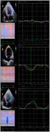
Background: The complete left bundle branch block (CLBBB) results in ventricular dyssynchrony and a reduction in systolic and diastolic efficiency. We noticed a distinct clockwise rotation of the left ventricle (LV) in patients with CLBBB ("longitudinal rotation").
Aim: The aim of this study was to quantify the "longitudinal rotation" of the LV in patients with CLBBB in comparison to patients with normal conduction or complete right bundle branch block (CRBBB).
Methods: Sixty consecutive patients with normal QRS, CRBBB, or CLBBB were included. Stored raw data DICOM 2D apical-4 chambers view images cine clips were analyzed using EchoPac plugin version 203 (GE Vingmed Ultrasound AS, Horten, Norway). In EchoPac-Q-Analysis, 2D strain application was selected. Instead of apical view algorithms, the SAX-MV (short axis-mitral valve level) algorithm was selected for analysis. A closed loop endocardial contour was drawn to initiate the analysis. The "posterior" segment (representing the mitral valve) was excluded before finalizing the analysis. Longitudinal rotation direction, peak angle, and time-to-peak rotation were recorded.
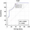
Results: All patients with CLBBB (n = 21) had clockwise longitudinal rotation with mean four chamber peak rotation angle of -3.9 ± 2.4°. This rotation is significantly larger than in patients with normal QRS (-1.4 ± 3°, p = 0.005) and CRBBB (0.1 ± 2.2°, p = 0.00001). Clockwise rotation was found to be correlated to QRS duration in patients with the non-RBBB pattern. The angle of rotation was not associated with a lower ejection fraction or the presence of regional wall abnormalities.
Conclusions: Significant clockwise longitudinal rotation was found in CLBBB patients compared to normal QRS or CRBBB patients using speckle-tracking echocardiography.
Keywords: CLBBB; clockwise; longitudinal; rotation.
Conflict of interest statement
The authors declare no conflict of interest.
Figures


References
-
- Bax J.J., Ansalone G., Breithardt O.A., Derumeaux G., Leclercq C., Schalij M.J., Sogaard P., John Sutton M.S., Petros Nihoyannopoulos MD, FRCP, FACC Echocardiographic evaluation of cardiac resynchronization therapy: Ready for routine clinical use? A critical appraisal. J. Am. Coll. Cardiol. 2004;44:1–9. doi: 10.1016/j.jacc.2004.02.055. - DOI - PubMed
-
- Pitzalis M.V., Iacoviello M., Romito R., Massari F., Rizzon B., Luzzi G., Guida P., Andriani A., Mastropasqua F., Rizzon P. Cardiac resynchronization therapy tailored by echocardiographic evaluation of ventricular asynchrony. J. Am. Coll. Cardiol. 2002;40:1615–1622. doi: 10.1016/S0735-1097(02)02337-9. - DOI - PubMed
-
- Yu C.-M., Fung J.W.-H., Zhang Q., Chan C.-K., Chan Y.-S., Lin H., Kum L.C., Kong S.-L., Zhang Y., Sanderson J.E., et al. Tissue Doppler Imaging Is Superior to Strain Rate Imaging and Postsystolic Shortening on the Prediction of Reverse Remodeling in Both Ischemic and Nonischemic Heart Failure After Cardiac Resynchronization Therapy. Circulation. 2004;110:66–73. doi: 10.1161/01.CIR.0000133276.45198.A5. - DOI - PubMed
-
- Yu C.-M., Chau E., Sanderson J.E., Fan K., Tang M.-O., Fung W.-H., Lin H., Kong S.-L., Lam Y.-M., Hill M.R., et al. Tissue Doppler Echocardiographic Evidence of Reverse Remodeling and Improved Synchronicity by Simultaneously Delaying Regional Contraction After Biventricular Pacing Therapy in Heart Failure. Circulation. 2002;105:438–445. doi: 10.1161/hc0402.102623. - DOI - PubMed
LinkOut - more resources
Full Text Sources
Research Materials

