Case Reports
. 2023 Jan 25;12(3):938.
doi: 10.3390/jcm12030938.

Jacob's Disease: Case Series, Extensive Literature Review and Classification Proposal

Affiliations
Case Reports

Jacob's Disease: Case Series, Extensive Literature Review and Classification Proposal

Luca Raccampo et al. J Clin Med. .

Abstract

Jacob's disease is a rare entity consisting of the formation of a pseudojoint between an abnormal coronoid process of the mandible and the inner surface of the zygomatic bone. First described by Jacob in 1899, its diagnosis and definition have never been entirely univocal. In this paper, we present three emblematic cases and an extensive review of the literature on Jacob's disease. Given the variability observed in the presentation of the disease, we have developed a proposal for the classification, here reported.

Keywords: Jacob’s disease; coronoid hyperplasia; osteochondroma; temporomandibular surgery.


Conflict of interest statement

The authors declare no conflict of interest.

Figures

Figure 1
Figure 1
Patient’s OPG showing no evident anomalies.
Figure 2
Figure 2
Cinematic 3D reconstruction (Mimics, Materialise NV, Leuven, Belgium) showing the pseudojoint established between the altered right CPM and the inner aspect of zygomatic bone.
Figure 3
Figure 3
SPET-CT showing bone reworking in correspondence to the joint between the jaw and the right zygomatic bone compared to the contralateral homologous site.
Figure 4
Figure 4
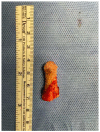
Right mushroom-shaped coronoid process excised.
Figure 5
Figure 5
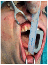
Patient showing an MMO of 20 mm and a slight deviation to the right at first physical examination.
Figure 6
Figure 6
Cinematic 3D reconstruction (Mimics, Materialise NV., Leuven, Belgium) showing the established right JD.
Figure 7
Figure 7
3D CT reconstructions showing an enlarged left CPM.
Figure 8
Figure 8
Surgical sequence of the intraoral coronoidectomy. The patient shows an immediate improvement in the MMO up to 40 mm.
Figure 9
Figure 9
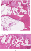
Hematoxylin–eosin staining of the lesion. Sections of compact, trabecular bone tissue covered by a layer of hyaline cartilage and fibrous cartilage overlaid by dense fibrous connective tissue (a,b).
Figure 10
Figure 10
MMO of 20 mm at first clinical examination.
Figure 11
Figure 11
3-dimensional dynamic reconstruction showing the bilateral pseudojoint determining a bilateral JD.
Figure 12
Figure 12
MMO of 50 mm right after bilateral coronoidectomy.

References

    1. Jacob O. Une cause rare de constriction permanente des machoires. Bull. Mem. Soc. Anat. Paris. 1899;1:917.
    1. Langenbeck B. Angeborene Kleinert der Unterkiefer. Langenbecks Arch. 1861;1:451.
    1. McLoughlin P., Hopper C., Bowley N. Hyperplasia of the mandibular coronoid process: An analysis of 31 cases and a review of the literature. J. Oral Maxillofac. Surg. 1995;53:250–255. doi: 10.1016/0278-2391(95)90219-8. - DOI - PubMed
    1. Emekli U., Aslan A., Onel D., Cizmeci O., Demiryont M. Osteochondroma of the coronoid process (Jacob’s disease) J. Oral Maxillofac. Surg. 2002;60:1354–1356. doi: 10.1053/joms.2002.35742. - DOI - PubMed
    1. Patel R., Obeid G. Osteochondroma of the Zygomatic Arch: A Case Report and Review of the Literature. J. Oral Maxillofac. Surg. 2018;76:1912–1916. doi: 10.1016/j.joms.2018.03.038. - DOI - PubMed

Publication types

LinkOut - more resources