. 2023 Jan 28;12(3):999.
doi: 10.3390/jcm12030999.

GLI1 Deficiency Impairs the Tendon-Bone Healing after Anterior Cruciate Ligament Reconstruction: In Vivo Study Using Gli1-Transgenic Mice

Affiliations

GLI1 Deficiency Impairs the Tendon-Bone Healing after Anterior Cruciate Ligament Reconstruction: In Vivo Study Using Gli1-Transgenic Mice

Yake Liu et al. J Clin Med. .

Abstract

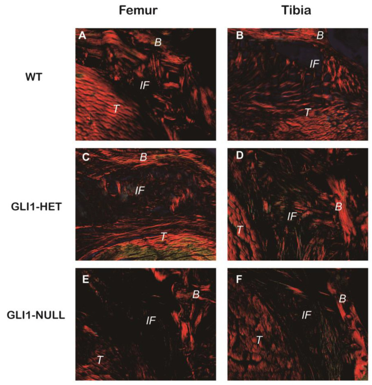
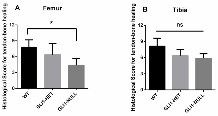
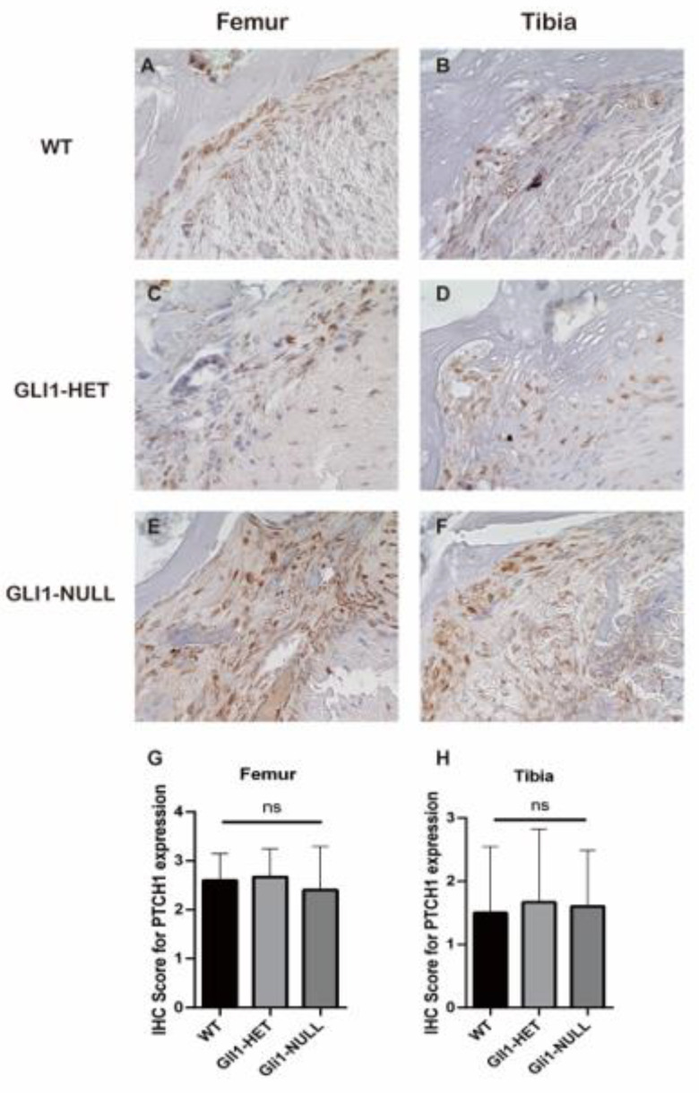
Hedgehog (Hh) signaling plays a fundamental role in the enthesis formation process and GLI-Kruppel family member GLI1 (Gli1) is a key downstream mediator. However, the role of Gli1 in tendon-bone healing after anterior cruciate ligament reconstruction (ACLR) is unknown. To evaluate the tendon-bone healing after ACLR in Gli1LacZ/LacZ (GLI1-NULL) mice, and compare Gli1LacZ/WT (GLI1-HET) and Gli1WT/WT wild type (WT) mice, a total of 45 mice, 15 mice each of GLI1-NULL, GLI1-HET and WT were used in this study. All mice underwent microsurgical ACLR at 12 weeks of age. Mice were euthanized at 4 weeks after surgery and were used for biomechanical testing, histological evaluation, and micro-CT analysis. The GLI1-NULL group had significantly lower biomechanical failure force, poorer histological healing, and lower BV/TV when compared with the WT and GLI1-HET groups. These significant differences were only observed at the femoral tunnel. Immunohistology staining showed positive expression of Indian hedgehog (IHH) and Patched 1(PTCH1) in all three groups, which indicated the activation of the Hh signal pathway. The GLI1 was negative in the GLI1-NULL group, validating the absence of GLI1 protein in these mice. These results proved that activation of the Hh signaling pathway occurs during ACL graft healing, and the function of Gli1 was necessary for tendon-bone healing. Healing in the femoral tunnel is more obviously impaired by Gli1 deficiency. Our findings provide further insight into the molecular mechanism of tendon-bone healing and suggest that Gli1 might represent a novel therapeutic target to improve tendon-bone healing after ACLR.

Keywords: Gli1; anterior cruciate ligament reconstruction; hedgehog; tendon–bone healing.


Conflict of interest statement

The authors declare no conflict of interest.

Figures

Figure 1
Figure 1
Study design.
Figure 2
Figure 2
Biomechanical testing results of wild-type, GLI1-HET and GLI1-NULL mice at 4 weeks after ACLR. (A) The failure force in the GLI1-NULL group was significantly lower than the GLI1-HET or WT counterparts at 4 weeks after surgery. (B) Stiffness was comparable among groups. Values are presented as mean ± SD. *, p < 0.05.
Figure 3
Figure 3
Hematoxylin and eosin staining of the tendon–bone interface of wild-type (A,B), GLI1-HET (C,D), and GLI1-NULL (E,F) mice at 4 weeks after ACLR (20×). Obvious fibrovascular tissue and wider and more distinct interface in the tendon–bone connection at both femoral and tibial sides could be observed in the GLI1-NULL group. (B: Bone tissue, IF: Interface, T: Tendon graft).
Figure 4
Figure 4
Safranin-O staining of the tendon–bone interface of wild-type (A,B), GLI1-HET (C,D) and GLI1-NULL (E,F) mice at 4 weeks after ACLR (40×). Fibrocartilage-like cells were observed at the tibial tunnel interface in the WT group and Gli1-HET group. (B: Bone tissue, IF: Interface, T: Tendon graft, *: Fibrocartilage-like cell).
Figure 5
Figure 5
Picrosirius red staining of the tendon–bone interface of Wild-Type (A,B), GLI1-HET (C,D), and GLI1-NULL (E,F) mice at 4 weeks after ACLR (40×). Oblique or perpendicular collagen fibers that crossed the tendon–bone interface were formed in the WT group and GLI1-HET group, while there were minimal, poorly organized connecting collagen fibers at the interface in the GLI1-NULL group. (B: Bone tissue, IF: Interface, T: Tendon graft).
Figure 6
Figure 6
Histologic scores for tendon–bone healing at femoral (A) and tibial (B) sides in wild-type, GLI1-HET and GLI1-NULL mice at 4 weeks after ACLR. Values are presented as mean ± SD. The WT group had a significantly higher score on the femoral side when compared with the GLI1-NULL group (p = 0.04).
Figure 7
Figure 7
Representative images of IHH immunohistological staining of the tendon–bone interface of wild-type (A,B), GLI1-HET (C,D) and GLI1-NULL (E,F) mice at 4 weeks after ACLR (40×). All three groups had positive IHH expression, with no significant difference among groups (G,H). Values are presented as mean ± SD.
Figure 8
Figure 8
Representative images of PTCH1 immunohistological staining of the tendon–bone interface of wild-type (A,B), GLI1-HET (C,D) and GLI1-NULL (E,F) mice at 4 weeks after ACLR (40×). All three groups had positive PTCH1 expression, with no significant difference among groups (G,H). Values are presented as mean ± SD.
Figure 9
Figure 9
Representative images of GLI1 immunohistological staining of the tendon–bone interface of wild-type (A,B), GLI1-HET (C,D) and GLI1-NULL (E,F) mice at 4 weeks after ACLR (40×). GLI1 was positive at the interface only in the WT and GLI1-HET groups and was negative in the GLI1-NULL group. This difference was significant on the femoral side. (G,H) Values are presented as mean ± SD. **, p < 0.01.
Figure 10
Figure 10
Results of micro-CT analyses for wild-type, GLI1-HET, and GLI1-NULL mice at 4 weeks after ACLR (40×). (A,B) Bone volume/total volume (BV/TV) in all three groups. Values are presented as mean ± SD. *, p < 0.05. (C) 3D-reconstruction images of all three groups (E: tunnel exit, M: mid-tunnel, A: aperture).

References

    1. Grassi A., Carulli C., Innocenti M., Mosca M., Zaffagnini S., Bait C., SIGASCOT Arthroscopy Committee New Trends in Anterior Cruciate Ligament Reconstruction: A Systematic Review of National Surveys of the Last 5 Years. Joints. 2018;6:177–187. doi: 10.1055/s-0038-1672157. - DOI - PMC - PubMed
    1. Webster K.E., Feller J.A., Hameister K.A. Bone tunnel enlargement following anterior cruciate ligament reconstruction: A randomised comparison of hamstring and patellar tendon grafts with 2-year follow-up. Knee Surg. Sports Traumatol. Arthrosc. 2001;9:86–91. doi: 10.1007/s001670100191. - DOI - PubMed
    1. Nebelung W., Becker R., Urbach D., Röpke M., Roessner A. Histological findings of tendon-bone healing following anterior cruciate ligament reconstruction with hamstring grafts. Arch. Orthop. Trauma Surg. 2003;123:158–163. doi: 10.1007/s00402-002-0463-y. - DOI - PubMed
    1. Rodeo S.A., Arnoczky S.P., Torzilli P.A., Hidaka C., Warren R.F. Tendon-healing in a bone tunnel. A biomechanical and histological study in the dog. J. Bone Joint Surg. Am. 1993;75:1795–1803. doi: 10.2106/00004623-199312000-00009. - DOI - PubMed
    1. Benjamin M., McGonagle D. Entheses: Tendon and ligament attachment sites. Scand. J. Med. Sci. Sports. 2009;19:520–527. doi: 10.1111/j.1600-0838.2009.00906.x. - DOI - PubMed

LinkOut - more resources