

Ultrasound during the COVID-19 Pandemic: A Global Approach
- PMID: 36769702
- PMCID: PMC9918296
- DOI: 10.3390/jcm12031057
Ultrasound during the COVID-19 Pandemic: A Global Approach
Abstract
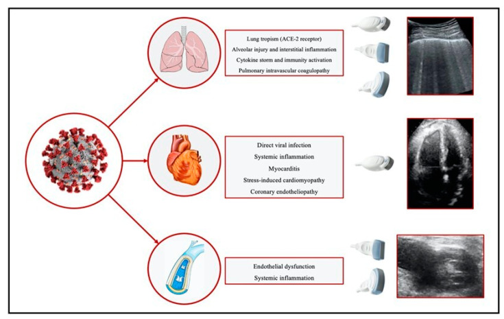
SARS-CoV-2 (severe acute respiratory syndrome Coronavirus-2) rapidly spread worldwide as COVID-19 (Coronavirus disease 2019), causing a costly and deadly pandemic. Different pulmonary manifestations represent this syndrome's most common clinical manifestations, together with the cardiovascular complications frequently observed in these patients. Ultrasound (US) evaluations of the lungs, heart, and lower limbs may be helpful in the diagnosis, follow-up, and prognosis of patients with COVID-19. Moreover, POCUS (point-of-care ultrasound) protocols are particularly useful for patients admitted to intensive care units. The present review aimed to highlight the clinical conditions during the SARS-CoV-2 pandemic in which the US represents a crucial diagnostic tool.
Keywords: COVID-19; echocardiography; intensive care unit; lung ultrasound; ultrasound.
Conflict of interest statement
The authors declare no conflict of interest.
Figures
References
-
- D’Andrea A., Mele D., Palermi S., Lombardi A., Di Giannuario G., Rizzo M., Campana M., Marrazzo G., Scarafile R., Gimelli A., et al. Ecografia del polmone: Quello che il cardiologo dovrebbe conoscere. G. Ital. di Cardiol. 2021;22:638–647. - PubMed
-
- Bajema K.L., Oster A.M., McGovern O.L., Lindstrom S., Stenger M.R., Anderson T.C., Isenhour C., Clarke K.R., Evans M.E., Chu V.T., et al. Persons Evaluated for 2019 Novel Coronavirus-United States, January 2020. MMWR Morb. Mortal Wkly. Rep. 2020;69:166–170. doi: 10.15585/mmwr.mm6906e1. - DOI - PMC - PubMed
Publication types
LinkOut - more resources
Full Text Sources
Miscellaneous
