Effect of Hydrogen Peroxide on the Surface and Attractiveness of Various Zirconia Implant Materials on Human Osteoblasts: An In Vitro Study
- PMID: 36769968
- PMCID: PMC9918077
- DOI: 10.3390/ma16030961
Effect of Hydrogen Peroxide on the Surface and Attractiveness of Various Zirconia Implant Materials on Human Osteoblasts: An In Vitro Study
Abstract
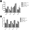
The aim of this in vitro study was to investigate the effect of hydrogen peroxide (H2O2) on the surface properties of various zirconia-based dental implant materials and the response of human alveolar bone osteoblasts. For this purpose, discs of two zirconia-based materials with smooth and roughened surfaces were immersed in 20% H2O2 for two hours. Scanning electron and atomic force microscopy showed no topographic changes after H2O2-treatment. Contact angle measurements (1), X-ray photoelectron spectroscopy (2) and X-ray diffraction (3) indicated that H2O2-treated surfaces (1) increased in hydrophilicity (p < 0.05) and (2) on three surfaces the carbon content decreased (33-60%), while (3) the monoclinic phase increased on all surfaces. Immunofluorescence analysis of the cell area and DNA-quantification and alkaline phosphatase activity revealed no effect of H2O2-treatment on cell behavior. Proliferation activity was significantly higher on three of the four untreated surfaces, especially on the smooth surfaces (p < 0.05). Within the limitations of this study, it can be concluded that exposure of zirconia surfaces to 20% H2O2 for 2 h increases the wettability of the surfaces, but also seems to increase the monoclinic phase, especially on roughened surfaces, which can be considered detrimental to material stability. Moreover, the H2O2-treatment has no influence on osteoblast behavior.
Keywords: cell culture; dental implant surface; hydrogen peroxide; osseointegration; primary human alveolar bone-derived osteoblasts; zirconia.
Conflict of interest statement
The authors declare no conflict of interest.
Figures


Similar articles
-
Effect of ultraviolet photofunctionalisation on the cell attractiveness of zirconia implant materials.Eur Cell Mater. 2015 Jan 23;29:82-94; discussion 95-6. doi: 10.22203/ecm.v029a07. Eur Cell Mater. 2015. PMID: 25612543
-
Influence of ultraviolet photofunctionalization on the surface characteristics of zirconia-based dental implant materials.Dent Mater. 2015 Feb;31(2):e14-24. doi: 10.1016/j.dental.2014.10.008. Epub 2014 Nov 22. Dent Mater. 2015. PMID: 25467951
-
Human fetal osteoblast behavior on zirconia dental implants and zirconia disks with microstructured surfaces. An experimental in vitro study.Clin Oral Implants Res. 2016 Nov;27(11):e144-e153. doi: 10.1111/clr.12585. Epub 2015 Mar 25. Clin Oral Implants Res. 2016. PMID: 25809053
-
Titanium dental implant surfaces obtained by anodic spark deposition - From the past to the future.Mater Sci Eng C Mater Biol Appl. 2016 Dec 1;69:1429-41. doi: 10.1016/j.msec.2016.07.068. Epub 2016 Jul 26. Mater Sci Eng C Mater Biol Appl. 2016. PMID: 27612843 Review.
-
Zirconia surface modifications for implant dentistry.Mater Sci Eng C Mater Biol Appl. 2019 May;98:1294-1305. doi: 10.1016/j.msec.2019.01.062. Epub 2019 Jan 16. Mater Sci Eng C Mater Biol Appl. 2019. PMID: 30813009 Free PMC article. Review.
Cited by
-
Efficacy of a Solution Containing 33% Trichloroacetic Acid and Hydrogen Peroxide in Decontaminating Machined vs. Sand-Blasted Acid-Etched Titanium Surfaces.J Funct Biomater. 2024 Jan 12;15(1):21. doi: 10.3390/jfb15010021. J Funct Biomater. 2024. PMID: 38248688 Free PMC article.
-
Implant Surface Decontamination Methods That Can Impact Implant Wettability.Materials (Basel). 2024 Dec 20;17(24):6249. doi: 10.3390/ma17246249. Materials (Basel). 2024. PMID: 39769848 Free PMC article. Review.
-
Zirconia in Dental Implantology: A Review of the Literature with Recent Updates.Bioengineering (Basel). 2025 May 19;12(5):543. doi: 10.3390/bioengineering12050543. Bioengineering (Basel). 2025. PMID: 40428162 Free PMC article. Review.
References
LinkOut - more resources
Full Text Sources

