

Microfluidic Fabrication of Gadolinium-Doped Hydroxyapatite for Theragnostic Applications
- PMID: 36770462
- PMCID: PMC9921701
- DOI: 10.3390/nano13030501
Microfluidic Fabrication of Gadolinium-Doped Hydroxyapatite for Theragnostic Applications
Abstract
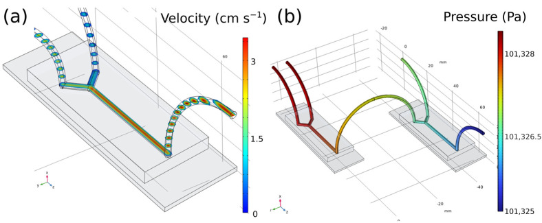
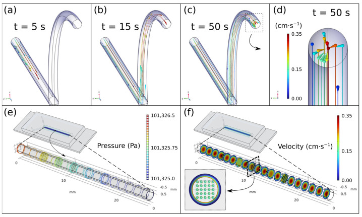
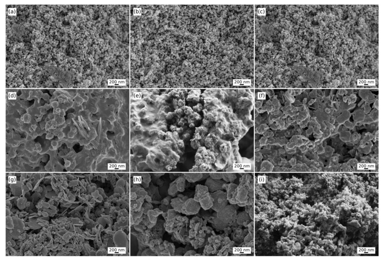
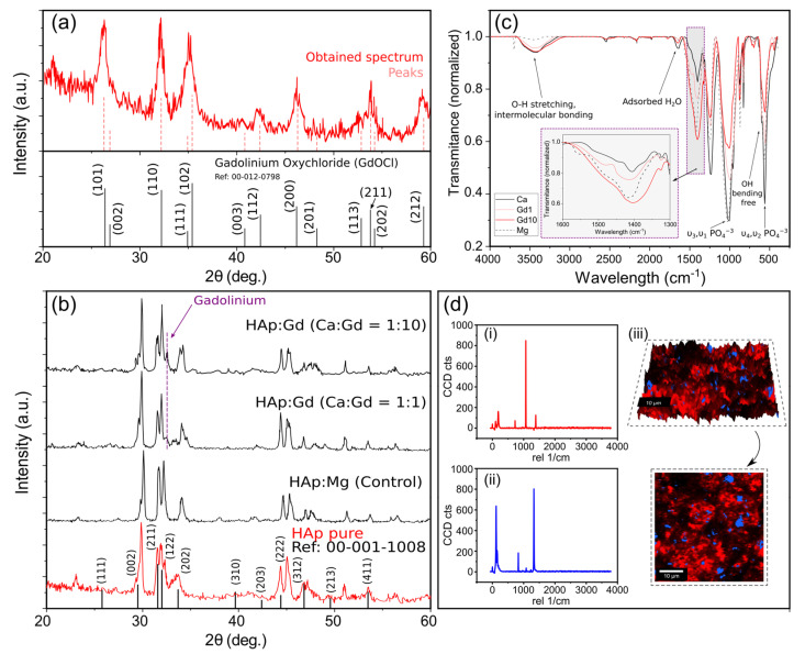
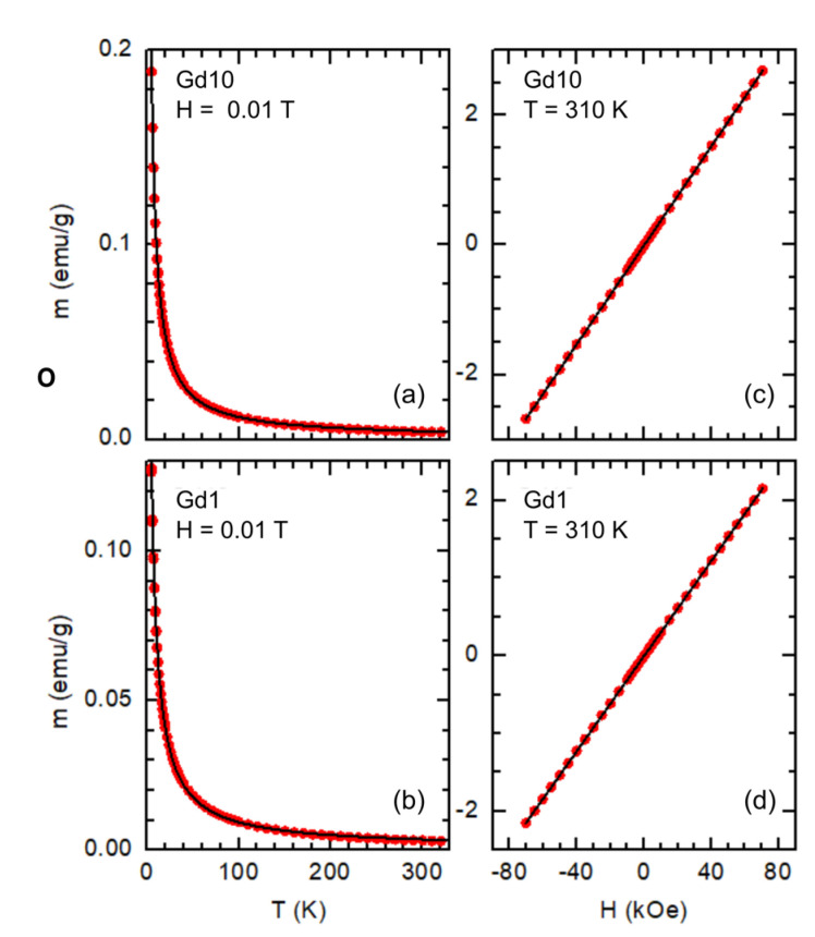
Among the several possible uses of nanoparticulated systems in biomedicine, their potential as theragnostic agents has received significant interest in recent times. In this work, we have taken advantage of the medical applications of Gadolinium as a contrast agent with the versatility and huge array of possibilities that microfluidics can help to create doped Hydroxyapatite nanoparticles with magnetic properties in an efficient and functional way. First, with the help of Computational Fluid Dynamics (CFD), we performed a complete and precise study of all the elements and phases of our device to guarantee that our microfluidic system worked in the laminar regime and was not affected by the presence of nanoparticles through the flow requisite that is essential to guarantee homogeneous diffusion between the elements or phases in play. Then the obtained biomaterials were physiochemically characterized by means of XRD, FE-SEM, EDX, confocal Raman microscopy, and FT-IR, confirming the successful incorporation of the lanthanide element Gadolinium in part of the Ca (II) binding sites. Finally, the magnetic characterization confirmed the paramagnetic behaviour of the nanoparticles, demonstrating that, with a simple and automatized system, it is possible to obtain advanced nanomaterials that can offer a promising and innovative solution in theragnostic applications.
Keywords: Computational Fluid Dynamics (CFD); microfluidics; nanomaterials; theragnostic; tissue engineering.
Conflict of interest statement
The authors declare no conflict of interest.
Figures
Similar articles
-
Single-Stage Microfluidic Synthesis Route for BaGdF5:Tb3+-Based Nanocomposite Materials: Synthesis, Characterization and Biodistribution.Int J Mol Sci. 2023 Dec 5;24(24):17159. doi: 10.3390/ijms242417159. Int J Mol Sci. 2023. PMID: 38138988 Free PMC article.
-
Facile synthesis and functionalization of color-tunable Ln3+-doped KGdF4 nanoparticles on a microfluidic platform.Mater Sci Eng C Mater Biol Appl. 2020 Mar;108:110381. doi: 10.1016/j.msec.2019.110381. Epub 2019 Nov 1. Mater Sci Eng C Mater Biol Appl. 2020. PMID: 31924035
-
Lanthanide-Activated Nanoparticles: A Toolbox for Bioimaging, Therapeutics, and Neuromodulation.Acc Chem Res. 2020 Nov 17;53(11):2692-2704. doi: 10.1021/acs.accounts.0c00513. Epub 2020 Oct 26. Acc Chem Res. 2020. PMID: 33103883 Review.
-
Noble microfluidic system for bioceramic nanoparticles engineering.Mater Sci Eng C Mater Biol Appl. 2019 Sep;102:221-227. doi: 10.1016/j.msec.2019.04.037. Epub 2019 Apr 13. Mater Sci Eng C Mater Biol Appl. 2019. PMID: 31146994
-
Microfluidic Platforms toward Rational Material Fabrication for Biomedical Applications.Small. 2020 Mar;16(9):e1903798. doi: 10.1002/smll.201903798. Epub 2019 Oct 25. Small. 2020. PMID: 31650698 Review.
Cited by
-
Synthesis and Characterization of Hydrogel Droplets Containing Magnetic Nano Particles, in a Microfluidic Flow-Focusing Chip.Gels. 2023 Jun 19;9(6):501. doi: 10.3390/gels9060501. Gels. 2023. PMID: 37367170 Free PMC article.
-
Exploring Gd3+-activated calcium-based host materials for phototherapy lamps: A comprehensive review.Heliyon. 2024 Jul 11;10(15):e34477. doi: 10.1016/j.heliyon.2024.e34477. eCollection 2024 Aug 15. Heliyon. 2024. PMID: 39157368 Free PMC article. Review.
-
In Vivo Assessment of High-Strength and Corrosion-Controlled Magnesium-Based Bone Implants.Bioengineering (Basel). 2023 Jul 24;10(7):877. doi: 10.3390/bioengineering10070877. Bioengineering (Basel). 2023. PMID: 37508904 Free PMC article.
References
-
- Wang Z.L., Zhu G., Yang Y., Wang S., Pan C. Progress in nanogenerators for portable electronics. Mater. Today. 2012;15:532–543. doi: 10.1016/S1369-7021(13)70011-7. - DOI
LinkOut - more resources
Full Text Sources
Miscellaneous
