. 2023 Jan 18;28(3):970.
doi: 10.3390/molecules28030970.

DNA Base Excision Repair Intermediates Influence Duplex-Quadruplex Equilibrium

Affiliations

DNA Base Excision Repair Intermediates Influence Duplex-Quadruplex Equilibrium

Mark L Sowers et al. Molecules. .

Abstract

Although genomic DNA is predominantly duplex under physiological conditions, particular sequence motifs can favor the formation of alternative secondary structures, including the G-quadruplex. These structures can exist within gene promoters, telomeric DNA, and regions of the genome frequently found altered in human cancers. DNA is also subject to hydrolytic and oxidative damage, and its local structure can influence the type of damage and its magnitude. Although the repair of endogenous DNA damage by the base excision repair (BER) pathway has been extensively studied in duplex DNA, substantially less is known about repair in non-duplex DNA structures. Therefore, we wanted to better understand the effect of DNA damage and repair on quadruplex structure. We first examined the effect of placing pyrimidine damage products uracil, 5-hydroxymethyluracil, the chemotherapy agent 5-fluorouracil, and an abasic site into the loop region of a 22-base telomeric repeat sequence known to form a G-quadruplex. Quadruplex formation was unaffected by these analogs. However, the activity of the BER enzymes were negatively impacted. Uracil DNA glycosylase (UDG) and single-strand selective monofunctional uracil DNA glycosylase (SMUG1) were inhibited, and apurinic/apyrimidinic endonuclease 1 (APE1) activity was completely blocked. Interestingly, when we performed studies placing DNA repair intermediates into the strand opposite the quadruplex, we found that they destabilized the duplex and promoted quadruplex formation. We propose that while duplex is the preferred configuration, there is kinetic conversion between duplex and quadruplex. This is supported by our studies using a quadruplex stabilizing molecule, pyridostatin, that is able to promote quadruplex formation starting from duplex DNA. Our results suggest how DNA damage and repair intermediates can alter duplex-quadruplex equilibrium.

Keywords: DNA quadruplex; base excision repair; duplex-quadruplex equilibrium; glycosylase; pyridostatin; telomere.


Conflict of interest statement

The authors declare no conflict of interest.

Figures

Figure 1
Figure 1
Sequences of oligonucleotides used in this study. Synthetic oligonucleotides of the telomere repeat sequence (Tel22), known to form a quadruplex structure, were prepared with various modifications. CD studies were performed on oligos without a fluorophore or quencher [Oligos 1–2]. FRET studies used a 6-carboxyfluorescein (FAM) and a 3′-non-fluorescent quencher (BHQ1) [Oligos 3–4], containing thymine (T) or its analogs uracil (U), 5-hydroxymethyluracil (5hmU), 5-fluorouracil (5FU), and a stable synthetic abasic site (THF). For gel-based studies, FAM-only oligos were used [Oligos 5–6]. A non-quadruplex (NQ) sequence of the same length [Oligos 2 and 6] was also prepared. In certain experiments, the quadruplex strand was annealed to a complementary strand [Oligos 7–9] to form a duplex where ‘Y’ is either T, U, THF, or two shorter oligos that simulate a one-base gap. Created with Biorender.com.
Figure 2
Figure 2
Pyrimidine analogs in the loop region did not disrupt G-quadruplex formation. CD spectra were acquired for quadruplex Tel22-X oligonucleotides or a non-quadruplex-forming (NQ) oligonucleotide. A 4 μM solution was prepared in 20 mM Tris buffer, pH 7.4, 150 mM KCl, and 15 mM NaCl and CD spectra were obtained from 320 to 220 nm at 37 °C. The spectra here were consistent with the formation of a hybrid-type G-quadruplex [21]. The 5hmU-containing oligonucleotide appeared slightly different from the others likely due to both intra- and intermolecular hydrogen bonding of 5hmU and N7 of adjacent guanines [25]. Tel22-T and the NQ were titrated from 0 to 150 mM KCl in a 20 mM Tris buffer at pH 7.4. Using a one-site specific binding model in the PRISM software, we estimated Kd of 1.2 ± 0.2 mM for quadruplex binding while no changes in CD spectra were seen with the NQ oligonucleotide. Titration experiments were done in triplicate, error bars of S.D. were smaller than the data points. Oligonucleotides were equilibrated in buffer at RT for a minimum of 30 min.
Figure 3
Figure 3
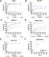
Quadruplex inhibited DNA glycosylase and APE1 activity. UDG, hSMUG1, and APE1 activity on quadruplex oligonucleotides (Tel22-X) containing U, 5hmU, 5FU, or THF was compared to duplex, or single-stranded DNA using the NQ-X oligonucleotides. (A) UDG (0.613 nM, 300:1 DNA to enzyme ratio) was fastest on single-stranded U (NQ-U) and duplex (U:A) followed by quadruplex (Tel22-U). (B) UDG (6.13 nM, 30:1 DNA to enzyme ratio) was overall less efficient on 5FU but the trend was the same, NQ-5FU > 5FU:A > Tel22-5FU. (C,D) hSMUG1 (6.2 nM, 30:1 DNA:enzyme ratio) followed a similar trend with either U or 5hmU. (E) Under the conditions tested, APE1 (1.31 nM, 140:1 DNA:enzyme ratio) was only active on duplex (THF:A) and no activity on Tel22-THF or NQ-THF was observed. (F) Because of the poor activity of APE1 on THF in a quadruplex, we wanted to estimate the rate of spontaneous β-elimination. We generated an abasic site in situ by using an excess of UDG to remove U from Tel22-U containing oligonucleotide. We then continued incubating the quadruplex containing abasic site for up to 50 h at 37 °C and measured spontaneous β-elimination over time using gel electrophoresis. The rate of elimination was determined by a linear fit to be 0.024 h−1 with a half-life of 21 h. Gels scans are shown in Figures S1–S16 Supplementary material.
Figure 4
Figure 4
G-quadruplex formation quenched fluorescence and the addition of a complementary strand promoted duplex formation. While quadruplexes are highly stable secondary DNA structures, in the presence of the complementary strand, they are in equilibrium with the corresponding duplex. A FRET strategy was employed to monitor quadruplex formation at 37 °C. Relatively low fluorescent excitation was observed when oligonucleotides formed a G-quadruplex as the fluorophore (FAM) and quencher (BHQ1) were in close contact. However, fluorescence increased when the quadruplex-containing sequence was annealed to its complementary strand to form a duplex (Inset). We then performed a time course experiment to examine the kinetics of quadruplex unfolding as it transitioned to a duplex. Black curves represent three independent experiments and the exponential fit in red. A single exponential fit poorly; therefore, we applied a double-exponential fit, which suggested that there was a relatively fast phase of quadruplex unfolding to one or more intermediate structures, followed by a slower complete unfolding to duplex. The first half-life was 1.9 ± 0.5 min and the second was 33 ± 11 min. The equation for the curve was y=A1(1ek1t)+A2(1ek2t) where k1 and k2 are 0.37 min−1 and 0.02 min−1, respectively. The first and second amplitudes were estimated to be 54.5 and 99.3, respectively.
Figure 5
Figure 5
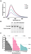
Reconstitution of repair intermediates promoted quadruplex formation. (A) Annealing Tel22-T with a complementary strand containing U (A:U) promoted duplex formation as indicated by an increase in fluorescence relative to the quadruplex-only control (Quad). When a complementary strand containing a stable abasic site (A:THF) was used instead, fluorescence was reduced relative to the A:U duplex, suggesting the destabilization of the duplex and promoting quadruplex. Interestingly, generating a gap in the complementary strand (A:Gap) further destabilized the duplex and shifted the equilibrium towards quadruplex. (B) Using a 20% native polyacrylamide gel, we demonstrated that A:U and A:THF were duplex as they migrated the same as the A:T control (Lanes 1–3). On the other hand, a gapped complement (A:Gap) showed some intermediate between duplex and quadruplex (Lane 4). Lane 5 was the quadruplex-only control. (C) We saw a concentration-dependent decrease in fluorescence when we titrated oligonucleotides that could form a duplex with a quadruplex-stabilizing small molecule, pyridostatin. Consistent with A and B, the A:Gap oligonucleotide had ~50% of the fluorescence as either A:U or A:THF. Oligonucleotides were prepared as a 1 μM solution in 20 mM Tris, pH 7.4, 150 mM KCl, and 15 mM NaCl and equilibrated in buffer for a minimum of 30 min. 1.2 equivalents of the corresponding complementary strands were annealed at 90 °C for 5 min and cooled at RT. Gel samples were prepared identically. Pyridostatin was allowed to equilibrate with oligonucleotides for 30 min at 37 °C and fluorescence emission of FAM was acquired using a qPCR instrument.
Figure 6
Figure 6
Base excision repair promotes quadruplex formation. In Figure 5, we demonstrated with synthetic oligonucleotides that the presence of a complementary strand containing an abasic site or gap destabilized the duplex and promoted quadruplex formation. Here, we simulated the same process but enzymatically prepared the abasic site and gapped DNA duplex using UDG or UDG and APE1, respectively. The fluorescent emission spectrum was taken for the quadruplex-only strand, duplex oligonucleotide containing a U in the C-rich strand (A:U), duplex [1 µM] incubated with 10 U of UDG (3.39 pmol, 34 nM), and duplex incubated with 10 U of UDG (3.39 pmol, 34 nM) and 20 U of APE1 (0.71 pmol, 7.1 nM) for 2 h at 37 °C in 100 µL total volume. Following UDG treatment, fluorescence decreased ~25% followed by a ~50% decrease in fluorescence with UDG and APE1. This suggested that the equilibrium between duplex and quadruplex could shift following DNA repair. We then wanted to estimate the time scale for secondary structure changes in the quadruplex strand, as a gap formed in the opposing strand. The duplex A:U containing oligonucleotide [1 µM] was treated with 1.25 U of UDG (0.42 pmol, 16.8 nM) and 2.5 U of APE1 (0.09 pmol, 3.6 nM) and fluorescence was monitored over time at 37 °C in a qPCR instrument in 25 µL total volume. As a gap was introduced into the opposing strand, fluorescence of the quadruplex strand decreased with time. This suggested that as the gap was introduced, the quadruplex-forming strand underwent relatively fast changes in configuration that brought the FAM and BHQ1 quencher closer together. The half-life was estimated to be 3.2 ± 0.4 min using a single exponential decay (Inset). This contrasted with the much slower unfolding of the quadruplex (Figure 4, Inset). The black curve represents the average of three replicates with the vertical lines as the S.D. The red curve shows a single exponential fit. In an identical experiment where the complementary oligonucleotide containing U was labeled with Cy5, we monitored the gap formation by gel (Figure S18).
Figure 7
Figure 7
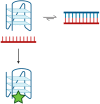
Duplex–quadruplex equilibrium scheme. Potentially quadruplex-forming regions of the genome are in equilibrium with duplex. Duplex is the preferred configuration in the presence of the complementary strand [22,23,24]. Transiently, a quadruplex may form that can be ‘trapped’ by quadruplex-binding ligands, such as pyridostatin (green star). The ligand stabilizes the quadruplex structure such that it prevents its unfolding and reforming duplex. Created with Biorender.com.

References

    1. Wallace S.S. Base Excision Repair: A Critical Player in Many Games. DNA Repair. 2014;19:14–26. doi: 10.1016/j.dnarep.2014.03.030. - DOI - PMC - PubMed
    1. Krokan H.E., Bjørås M. Base Excision Repair. Cold Spring Harb. Perspect. Biol. 2013;5:a012583. doi: 10.1101/cshperspect.a012583. - DOI - PMC - PubMed
    1. Wang G., Vasquez K.M. Impact of Alternative DNA Structures on DNA Damage, DNA Repair, and Genetic Instability. DNA Repair. 2014;19:143–151. doi: 10.1016/j.dnarep.2014.03.017. - DOI - PMC - PubMed
    1. Satange R., Chang C.K., Hou M.H. A Survey of Recent Unusual High-Resolution DNA Structures Provoked by Mismatches, Repeats and Ligand Binding. Nucleic Acids Res. 2018;46:6416–6434. doi: 10.1093/nar/gky561. - DOI - PMC - PubMed
    1. Bansal A., Kaushik S., Kukreti S. Non-Canonical DNA Structures: Diversity and Disease Association. Front. Genet. 2022;13:959258. doi: 10.3389/fgene.2022.959258. - DOI - PMC - PubMed