Potential Biological Properties of Lycopene in a Self-Emulsifying Drug Delivery System
- PMID: 36770886
- PMCID: PMC9920511
- DOI: 10.3390/molecules28031219
Potential Biological Properties of Lycopene in a Self-Emulsifying Drug Delivery System
Abstract
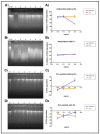
In recent years, lycopene has been highlighted due to its antioxidant and anti-inflammatory properties, associated with a beneficial effect on human health. The aim of this study was to advance the studies of antioxidant and anti-inflammatory mechanisms on human keratinocytes cells (HaCaT) of a self-emulsifying drug delivery system (SEDDS) loaded with lycopene purified from red guava (nanoLPG). The characteristics of nanoLPG were a hydrodynamic diameter of 205 nm, a polydispersity index of 0.21 and a zeta potential of -20.57, providing physical stability for the nanosystem. NanoLPG demonstrated antioxidant capacity, as shown using the ORAC methodology, and prevented DNA degradation (DNA agarose). Proinflammatory activity was evaluated by quantifying the cytokines TNF-α, IL-6 and IL-8, with only IL-8 showing a significant increase (p < 0.0001). NanoLPG showed greater inhibition of the tyrosinase and elastase enzymes, involved in the skin aging process, compared to purified lycopene (LPG). In vitro treatment for 24 h with 5.0 µg/mL of nanoLPG did not affect the viability of HaCaT cells. The ultrastructure of HaCaT cells demonstrated the maintenance of morphology. This contrasts with endoplasmic reticulum stresses and autophagic vacuoles when treated with LPG after stimulation or not with LPS. Therefore, the use of lycopene in a nanoemulsion may be beneficial in strategies and products associated with skin health.
Keywords: antioxidant; carotenoid; guava fruit; nanomedicine; skin care.
Conflict of interest statement
The authors declare no conflict of interest. The funders had no role in the design of the study; in the collection, analyses, or interpretation of data; in the writing of the manuscript, or in the decision to publish the results.
Figures


References
-
- Chiari-Andréo B.G., de Almeida-Cincotto M.G.J., Oshiro J.A., Jr., Taniguchi C.Y.Y., Chiavacci L.A., Isaac V.L.B. Nanoparticles for cosmetic use and its application. In: Grumezescu A.M., editor. Nanoparticles in Pharmacotherapy. Elsevier Inc.; Amsterdam, The Netherlands: 2019. pp. 113–146. - DOI
-
- Chuo S.C., Sepatar H.M. Application of nanotechnology for development of cosmetics. In: Setapsr S.H.M., Ahmad A., Jawaid M., editors. Nanotechnology for the Preparation of Cosmetis Using Plant-Based Extracts. Elsevier Inc.; Amsterdam, The Netherlands: 2022. pp. 327–344. - DOI
MeSH terms
Substances
Grants and funding
- 303557/2021-4/National Council for Scientific and Technological Development
- 0001/Coordenação de Aperfeicoamento de Pessoal de Nível Superior
- 01.08.0457.00/Financiadora de Estudos e Projetos
- 128959/2021-69/Foundation for Research Support of the Federal District
- UIDB/50016/2020/National Funds from FCT
LinkOut - more resources
Full Text Sources
Medical

