Review
. 2023 Jan 27;28(3):1231.
doi: 10.3390/molecules28031231.

Development and Application of Ruthenium(II) and Iridium(III) Based Complexes for Anion Sensing

Affiliations
Review

Development and Application of Ruthenium(II) and Iridium(III) Based Complexes for Anion Sensing

Ambreen Rashid et al. Molecules. .

Abstract

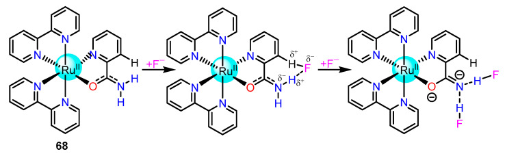
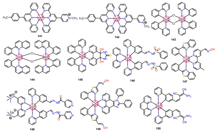
Improvements in the design of receptors for the detection and quantification of anions are desirable and ongoing in the field of anion chemistry, and remarkable progress has been made in this direction. In this regard, the development of luminescent chemosensors for sensing anions is an imperative and demanding sub-area in supramolecular chemistry. This decade, in particular, witnessed advancements in chemosensors based on ruthenium and iridium complexes for anion sensing by virtue of their modular synthesis and rich chemical and photophysical properties, such as visible excitation wavelength, high quantum efficiency, high luminescence intensity, long lifetimes of phosphorescence, and large Stokes shifts, etc. Thus, this review aims to summarize the recent advances in the development of ruthenium(II) and iridium(III)-based complexes for their application as luminescent chemosensors for anion sensing. In addition, the focus was devoted to designing aspects of polypyridyl complexes of these two transition metals with different recognition motifs, which upon interacting with different inorganic anions, produces desirable quantifiable outputs.

Keywords: anion sensing; luminescent chemosensors; ruthenium(II)/iridium(III) complexes.


Conflict of interest statement

The authors declare no conflict of interest.

Figures

Figure 1
Figure 1
Simplified Jablonski diagram of RuII/IrIII complexes showing triplet-state emission mechanism.
Figure 2
Figure 2
Structures of triazole-based Ru(II) complexes 110.
Figure 3
Figure 3
Structures of triazole-based Ru(II) complexes 1123.
Figure 4
Figure 4
X-ray crystal structure of H2PO4 adduct of 13 where supramolecular polymeric chain propagated via (a) C-I···O interaction through halogen bond; (b) combined C-I···O and π-π stacking interactions. [Adapted from reference [57]; Copyright © 2023, American Chemical Society].
Figure 5
Figure 5
Structures of biimidazole/imidazole-based Ru(II) complexes 2446 and the crystal structure of 24-NO3 complex. [Adapted from ccdc no.279202].
Figure 6
Figure 6
(a) Relative emission response of 36 (1.0 μM in pH = 7.00; 0.02 M HEPES buffer) at 593 nm in the presence of 5 mM potassium salts of the aforesaid anions; (b) Optimized structure of 36–CN at the B3LYP/6−31G* level for H, C, and N atoms and the SDD for Ru. [Adapted from reference [74]; Copyright © 2023, American Chemical Society]. (c) Emission color change under 365 nm UV lamp of 37–Cu2+ (10 μM) in the presence of 20 equiv. of different anions. [Adapted from reference [75]; © 2023 Elsevier B.V. All rights reserved].
Scheme 1
Scheme 1
Mode of hydrogen bonding interaction between 39 and CH3COO.
Scheme 2
Scheme 2
Mode of hydrogen bonding interaction between 42 and CH3COO.
Figure 7
Figure 7
(a) Photographs of acetonitrile solutions of 44 (10μM) taken under a UV−lamp in the absence and presence of 10 equiv. of anions. (b) The PL emission intensity ratios of 44 were plotted in the presence and absence of the anions (λex = 460 nm). [Reproduced from reference [81]; Copyright © 2023, Royal Society of Chemistry].
Figure 8
Figure 8
Structures of imidazole-based Ru(II) complexes 4754.
Figure 9
Figure 9
Structures of Amide/Sulphonamide/Picolinamide-based Ru(II) complexes 5570.
Scheme 3
Scheme 3
Modes of binding of 57 with CH3COO and CN.
Figure 10
Figure 10
(a)Two coordination modes of 2-picolinamide: (I) H2pia-k2N,O (II) Hpia-k2N,N’; (b) The naked eye color of the solution of only 68 (left), and after adding TBAF (right). [Adapted from reference [91]; © 2023 Elsevier B.V. All rights reserved].
Scheme 4
Scheme 4
The stepwise reaction of 68 with F to give di-F adduct.
Scheme 5
Scheme 5
Schematic representation of the formation of the 1:2 adduct of 69 and H2PO4.
Figure 11
Figure 11
Structures of dipyrrol-based Ru(II) complexes 7173.
Figure 12
Figure 12
Structures of urea-based Ru(II) complexes 7478.
Figure 13
Figure 13
Structures of Aldehyde group containing Ru(II) complexes 8184. (a) phosphorescence titration of 85 with CN (0.3 eq/time) (inset left: 85, right: 85 + 3CN). (b) Job′s plots of CN adduct complexes of 85. [Adapted from reference [109]; Copyright © 2023, Royal Society of Chemistry].
Figure 14
Figure 14
Structures of 2,2’-dipyridylamine−based Ru(II) complexes 8896 and (a) cyclic voltammograms of 9196, recorded in acetonitrile solution with Ag/AgCl/KCl(std.) electrode. [Adapted from reference [112]; © 2023 Elsevier B.V. All rights reserved].
Scheme 6
Scheme 6
Mechanism of deprotonation of 86 and 87 via hydrogen bonding interaction.
Scheme 7
Scheme 7
Formation of mono and di-F adduct of 97.
Figure 15
Figure 15
Structure of Ir(III) complex 98 and (a) naked eye color change; (b) emission color observed under a UV lamp in the absence and presence of F. [Adapted from reference [116]; Copyright © 2023, American Chemical Society].
Figure 16
Figure 16
Structures of Ir(III) complexes 99125.
Scheme 8
Scheme 8
Mechanism of PET process of 105 with “OFF-ON” F sensor.
Figure 17
Figure 17
Structures of Ir(III) complexes 126131; (a)The comparative response of complex 126 in UV−Vis, PL, and ECL upon the addition of anions. [Adapted from reference [129]; Copyright © 2023, Royal Society of Chemistry]. (b) luminescent on/off sensor for anions; (c) picture of 127 in chloroform and excess anions. [adapted from reference [130]; Copyright © 2023, Royal Society of Chemistry].
Figure 18
Figure 18
Structures of Ir(III) complexes 132135.
Figure 19
Figure 19
Structures of Ir(III) complexes 136137.
Figure 20
Figure 20
Structures of Ir(III) complexes 138140 and TEM images of nanoparticles found from anion titration of (a) Free 138 (b) 138 + 10 equiv. HPO42−; (c) 138 + 10 equiv. I; (d) 138 + 3 equiv. ClO4; (e) 138 + 5 equiv. ClO4; (f) 138 + 10 equiv. ClO4. [Adapted from reference [145]; Copyright © 2023, Royal Society of Chemistry].
Figure 21
Figure 21
Structures of Ir(III) complexes 141150.

Similar articles

Cited by

References

    1. Sessler J.L., Gale P.A., Cho W.S. Anion Receptor Chemistry. Royal Society of Chemistry; Cambridge, UK: 2006.
    1. Sessler J.L., Camiolo S., Gale P.A. Pyrrolic and polypyrrolic anion binding agents. Coord. Chem. Rev. 2003;240:17. doi: 10.1016/S0010-8545(03)00023-7. - DOI
    1. Kang S.O., Hossain M.A., Bowman-James K. Influence of dimensionality and charge on anion binding in amide-based macrocyclic receptors. Coord. Chem. Rev. 2006;250:3038–3052. doi: 10.1016/j.ccr.2006.06.006. - DOI
    1. Gale P.A., Quesada R. Anion coordination and anion-templated assembly: Highlights from 2002 to 2004. Coord. Chem. Rev. 2006;250:3219–3244. doi: 10.1016/j.ccr.2006.05.020. - DOI
    1. Gunnlaugsson T., Glynn M., Tocci G.M., Kruger P.E., Pfeffer F.M. Anion recognition and sensing in organic and aqueous media using luminescent and colorimetric sensors. Coord. Chem. Rev. 2006;250:3094–3117. doi: 10.1016/j.ccr.2006.08.017. - DOI

LinkOut - more resources