

Abnormal Maternal Body Mass Index and Customized Fetal Weight Charts: Improving the Identification of Small for Gestational Age Fetuses and Newborns
- PMID: 36771294
- PMCID: PMC9920601
- DOI: 10.3390/nu15030587
Abnormal Maternal Body Mass Index and Customized Fetal Weight Charts: Improving the Identification of Small for Gestational Age Fetuses and Newborns
Abstract
Background: Obesity and thinness are serious diseases, but cases with abnormal maternal weight have not been excluded from the calculations in the construction of customized fetal growth curves (CCs).
Method: To determine if the new CCs, built excluding mothers with an abnormal weight, are better than standard CCs at identifying SGA. A total of 16,122 neonates were identified as SGA, LGA, or AGA, using the two models. Logistic regression and analysis of covariance were used to calculate the OR and CI for adverse outcomes by group. Gestational age was considered as a covariable.
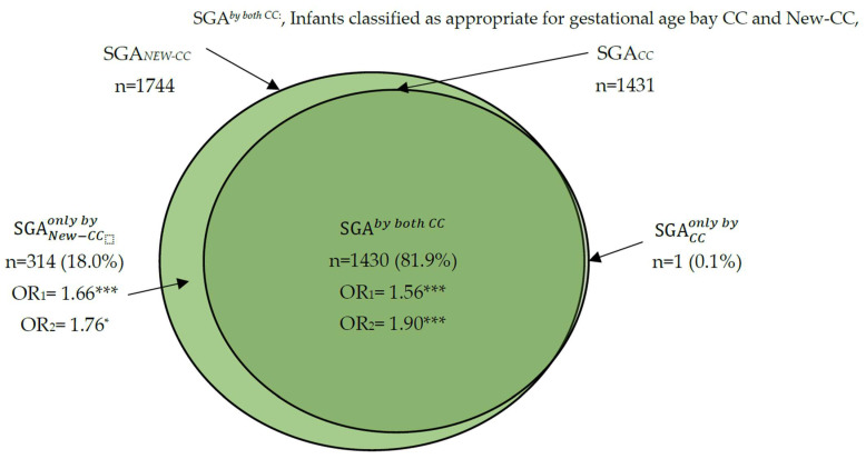
Results: The SGA rates by the new CCs and by the standard CCs were 11.8% and 9.7%, respectively. The SGA rate only by the new CCs was 18% and the SGA rate only by the standard CCs was 0.01%. Compared to AGA by both models, SGA by the new CCs had increased rates of cesarean section, (OR 1.53 (95% CI 1.19, 1.96)), prematurity (OR 2.84 (95% CI 2.09, 3.85)), NICU admission (OR 5.41 (95% CI 3.47, 8.43), and adverse outcomes (OR 1.76 (95% CI 1.06, 2.60). The strength of these associations decreased with gestational age.
Conclusion: The use of the new CCs allowed for a more accurate identification of SGA at risk of adverse perinatal outcomes as compared to the standard CCs.
Keywords: birthweight; customized growth charts; fetal weight; maternal body mass index; newborn weight; obesity; perinatal outcomes; small for gestational age; thinness.
Conflict of interest statement
The authors declare that the research was conducted in the absence of any commercial or financial relationships that could be construed as a potential conflict of interest.
Figures
Similar articles
-
Maternal Thinness and Obesity and Customized Fetal Weight Charts.Fetal Diagn Ther. 2021;48(7):551-559. doi: 10.1159/000515251. Epub 2021 Aug 18. Fetal Diagn Ther. 2021. PMID: 34407539
-
Derivation and assessment of a sex-specific fetal growth standard.J Matern Fetal Neonatal Med. 2022 Dec;35(25):9913-9921. doi: 10.1080/14767058.2022.2075696. Epub 2022 May 22. J Matern Fetal Neonatal Med. 2022. PMID: 35603475 Free PMC article.
-
"INTERGROWTH21st vs customized fetal growth curves in the assessment of the neonatal nutritional status: a retrospective cohort study of gestational diabetes".BMC Pregnancy Childbirth. 2020 Mar 4;20(1):139. doi: 10.1186/s12884-020-2845-y. BMC Pregnancy Childbirth. 2020. PMID: 32131758 Free PMC article.
-
Third-trimester uterine artery Doppler for prediction of adverse outcome in late small-for-gestational-age fetuses: systematic review and meta-analysis.Ultrasound Obstet Gynecol. 2020 May;55(5):575-585. doi: 10.1002/uog.21940. Ultrasound Obstet Gynecol. 2020. PMID: 31785172
-
[Review of the literature on intrauterine and birthweight charts].Gynecol Obstet Fertil Senol. 2023 May;51(5):256-269. doi: 10.1016/j.gofs.2022.09.014. Epub 2022 Oct 24. Gynecol Obstet Fertil Senol. 2023. PMID: 36302475 Review. French.
References
-
- Castro Conde J.R., González Campo C., González González N.L., Reyes Millan B., González Barrios D., Quintero Fuetes I. Assessment of neonatal EEG background and neurodevelopment in full-term small for their gestational age infants. Pediatr. Res. 2020;88:91–99. doi: 10.1038/s41390-019-0693-0. - DOI - PMC - PubMed
MeSH terms
LinkOut - more resources
Full Text Sources
Miscellaneous
