

Osteocytic Sclerostin Expression as an Indicator of Altered Bone Turnover
- PMID: 36771305
- PMCID: PMC9921466
- DOI: 10.3390/nu15030598
Osteocytic Sclerostin Expression as an Indicator of Altered Bone Turnover
Abstract
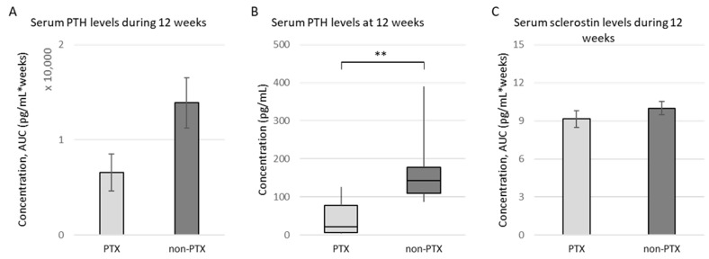
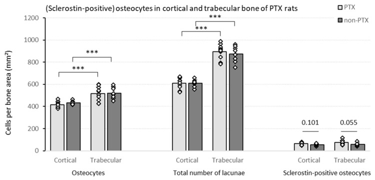
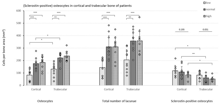
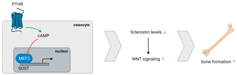
Renal osteodystrophy (ROD) is a complex and serious complication of chronic kidney disease (CKD), a major global health problem caused by loss of renal function. Currently, the gold standard to accurately diagnose ROD is based on quantitative histomorphometric analysis of trabecular bone. Although this analysis encompasses the evaluation of osteoblast and osteoclast number/activity, tfigurehe interest in osteocytes remains almost nihil. Nevertheless, this cell type is evidenced to perform a key role in bone turnover, particularly through its production of various bone proteins, such as sclerostin. In this study, we aim to investigate, in the context of ROD, to which extent an association exists between bone turnover and the abundance of osteocytes and osteocytic sclerostin expression in both the trabecular and cortical bone compartments. Additionally, the effect of parathyroid hormone (PTH) on bone sclerostin expression was examined in parathyroidectomized rats. Our results indicate that PTH exerts a direct inhibitory function on sclerostin, which in turn negatively affects bone turnover and mineralization. Moreover, this study emphasizes the functional differences between cortical and trabecular bone, as the number of (sclerostin-positive) osteocytes is dependent on the respective bone compartment. Finally, we evaluated the potential of sclerostin as a marker for CKD and found that the diagnostic performance of circulating sclerostin is limited and that changes in skeletal sclerostin expression occur more rapidly and more pronounced. The inclusion of osteocytic sclerostin expression and cortical bone analysis could be relevant when performing bone histomorphometric analysis for diagnostic purposes and to unravel pathological mechanisms of bone disease.
Keywords: bone turnover; chronic kidney disease (CKD); osteocyte; parathyroid hormone (PTH); parathyroidectomy (PTX); renal osteodystrophy (ROD); sclerostin.
Conflict of interest statement
The authors declare no conflict of interest.
Figures
Similar articles
-
Sclerostin, Osteocytes, and Wnt Signaling in Pediatric Renal Osteodystrophy.Nutrients. 2023 Sep 25;15(19):4127. doi: 10.3390/nu15194127. Nutrients. 2023. PMID: 37836411 Free PMC article.
-
FGF-23 and sclerostin in serum and bone of CKD patients.Clin Nephrol. 2023 May;99(5):209-218. doi: 10.5414/CN111111. Clin Nephrol. 2023. PMID: 36970967 Free PMC article.
-
Effects of dietary phosphate on adynamic bone disease in rats with chronic kidney disease--role of sclerostin?PLoS One. 2013 Nov 13;8(11):e79721. doi: 10.1371/journal.pone.0079721. eCollection 2013. PLoS One. 2013. PMID: 24236156 Free PMC article.
-
Sclerostin, Osteocytes, and Chronic Kidney Disease - Mineral Bone Disorder.Semin Dial. 2015 Nov-Dec;28(6):578-86. doi: 10.1111/sdi.12415. Epub 2015 Aug 19. Semin Dial. 2015. PMID: 26288182 Review.
-
Bone disease in pediatric chronic kidney disease.Pediatr Nephrol. 2013 Apr;28(4):569-76. doi: 10.1007/s00467-012-2324-4. Epub 2012 Oct 14. Pediatr Nephrol. 2013. PMID: 23064662 Free PMC article. Review.
Cited by
-
Evaluation of Bone Biomarkers in Renal Osteodystrophy.Life (Basel). 2024 Nov 25;14(12):1540. doi: 10.3390/life14121540. Life (Basel). 2024. PMID: 39768249 Free PMC article.
-
Sclerostin, Osteocytes, and Wnt Signaling in Pediatric Renal Osteodystrophy.Nutrients. 2023 Sep 25;15(19):4127. doi: 10.3390/nu15194127. Nutrients. 2023. PMID: 37836411 Free PMC article.
References
MeSH terms
Substances
LinkOut - more resources
Full Text Sources
Medical
