

Fruit Extract of Sechium chinantlense (Lira & F. Chiang) Induces Apoptosis in the Human Cervical Cancer HeLa Cell Line
- PMID: 36771372
- PMCID: PMC9920575
- DOI: 10.3390/nu15030667
Fruit Extract of Sechium chinantlense (Lira & F. Chiang) Induces Apoptosis in the Human Cervical Cancer HeLa Cell Line
Abstract
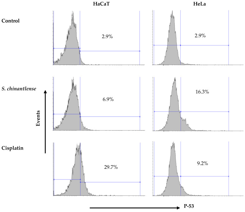
Sechium edule (Cucurbitaceae) is a commercial species of chayote and is just one of several species in the genus Sechium, whose extracts inhibit proliferation in tumor cell lines. The capacity of the wild species Sechium chinantlense (SCH) as an antitumor agent is unknown, as is the mechanism of action. In the present study, HeLa cervical cancer and HaCaT normal cell lines were treated with SCH and cell proliferation was inhibited in both cell lines in a dose-dependent manner similar to the effect of the antineoplastic agent cisplatin (Cis). Additionally, SCH arrested cell cycle progression but only in HeLa cells and induced apoptosis, as shown by phosphatidylserine translocation and caspase-3 activation, while Cis did so in both cell lines. Exploration of the mechanism of action of SCH in HeLa cells suggests that apoptosis was mediated by the intrinsic signaling pathway since there was no activation of caspase-8, but there was a release of cytochrome-c. These findings suggest that the SCH extract has the potential to selectively kill tumor cells by promoting apoptosis, without harming nontumor cells.
Keywords: apoptosis; cervical cancer; intrinsic signaling pathway.
Conflict of interest statement
The authors have no conflicts of interest to declare.
Figures


References
-
- The Global Cancer Observatory: Cancer Today. International Agency for Research on Cancer. [(accessed on 6 August 2022)]. Available online: https://gco.iarc.fr/today/fact-sheets-cancers.
-
- OMS (Organización Mundial de la Salud) Cáncer Cervicouterino. [(accessed on 6 August 2022)];2022 Available online: https://www.who.int/es/news-room/fact-sheets/detail/cervical-cancer.
-
- Cascales-Angosto M. Bases moleculares de la apoptosis. Anal. Real. Acad. Nal. Farm. 2003;69:36–64.
MeSH terms
Substances
Grants and funding
LinkOut - more resources
Full Text Sources
Research Materials
Miscellaneous
