. 2023 Jan 31;12(3):617.
doi: 10.3390/plants12030617.

An Interplay between Mitochondrial and ER Targeting of a Bacterial Signal Peptide in Plants

Affiliations

An Interplay between Mitochondrial and ER Targeting of a Bacterial Signal Peptide in Plants

Tatiana Spatola Rossi et al. Plants (Basel). .

Abstract

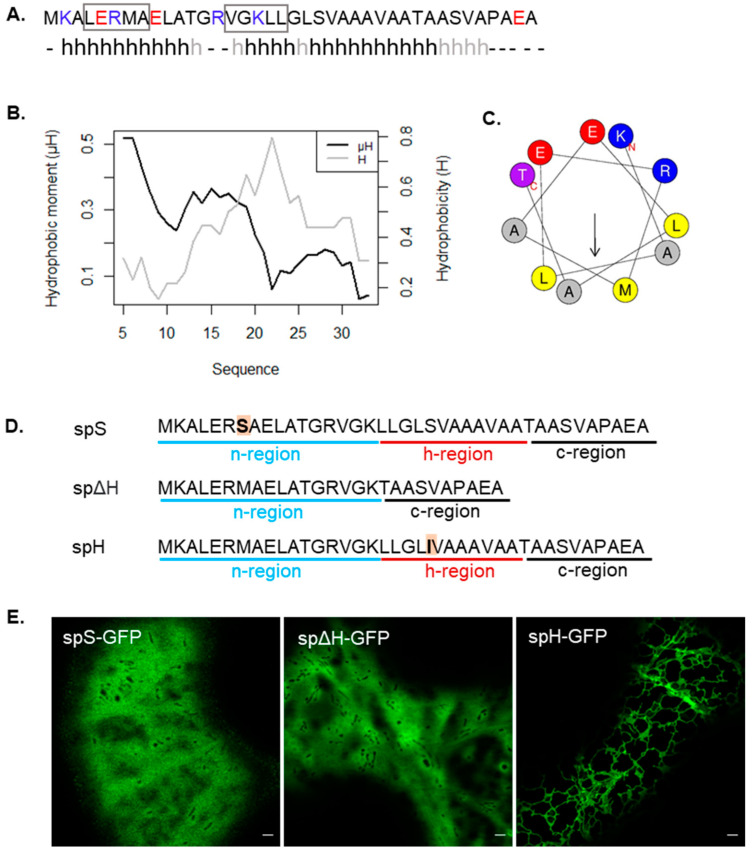
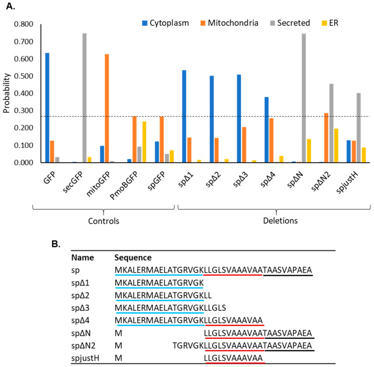
Protein targeting is essential in eukaryotic cells to maintain cell function and organelle identity. Signal peptides are a major type of targeting sequences containing a tripartite structure, which is conserved across all domains in life. They are frequently included in recombinant protein design in plants to increase yields by directing them to the endoplasmic reticulum (ER) or apoplast. The processing of bacterial signal peptides by plant cells is not well understood but could aid in the design of efficient heterologous expression systems. Here we analysed the signal peptide of the enzyme PmoB from methanotrophic bacteria. In plant cells, the PmoB signal peptide targeted proteins to both mitochondria and the ER. This dual localisation was still observed in a mutated version of the signal peptide sequence with enhanced mitochondrial targeting efficiency. Mitochondrial targeting was shown to be dependent on a hydrophobic region involved in transport to the ER. We, therefore, suggest that the dual localisation could be due to an ER-SURF pathway recently characterised in yeast. This work thus sheds light on the processing of bacterial signal peptides by plant cells and proposes a novel pathway for mitochondrial targeting in plants.

Keywords: ER-SURF; endoplasmic reticulum; mitochondria; plant cells; protein targeting; signal peptide; tobacco.


Conflict of interest statement

The authors declare no conflict of interest.

Figures

Figure 1
Figure 1
The PmoB signal peptide produces dual localisation to mitochondria and the ER in tobacco cells. (A) Diagram of PmoB (in grey) with two transmembrane domains (darker grey squares), comprising a signal peptide between residues 1–39 (black rectangle). n-, h- and c-regions are underlined in blue, red and black, respectively, and the signal peptidase cleavage site is highlighted in bold. GFP (green rectangle) was fused to the PmoB C-terminus. (B) Co-localisation of PmoB-GFP with mitochondria labelled with rhodamine B hexyl ester. (C) Co-localisation of PmoB-GFP with mitochondria at higher magnification. (D) Diagram of sp-GFP, comprising the PmoB signal peptide (black rectangle) fused upstream of GFP. (E) Co-localisation of sp-GFP with mitochondria label. (F) Co-localisation at higher magnification. (G) Co-localisation of sp-GFP with ER marker. (H) Schematic diagram for dual targeting and the ER-SURF pathway. For dual targeting, the signal peptide is recognized either by mitochondrial targeting factors or SRP and is targeted to both the mitochondria (Mito) and ER, respectively. A higher affinity for mitochondrial targeting factors confers a predominantly mitochondrial localisation (represented by the thicker arrow). In the ER-SURF pathway, the signal peptide first directs the protein to the ER surface, where chaperones (blue triangles) assist the subsequent funnelling to mitochondria (Mito). Scale bars = 5 μm.
Figure 2
Figure 2
Prediction of an N-terminal amphipathic helix in silico and signal peptide mutations impairing mitochondrial targeting. (A) Secondary structure prediction using Jpred 4 and PSIPRED 4.0. An h denotes helical conformation predicted by both software approaches, and light grey h prediction as helix only by PSIPRED 4.0. Basic amino acids are marked in blue, and acidic amino acids are marked in red. The two hydrophobic motifs predicted by MitoFates are highlighted using a rectangle. (B) Hydrophobic moment and hydrophobicity of 11 residue windows using HeliQuest. (C) Helical wheel diagram of the first 11 residues predicted as an α-helix showing a high hydrophobic moment. (D) Sequences of the signal peptide mutants generated. (E) Transient expression of mutants in tobacco cells with localisation of spS-GFP and spΔH-GFP to the cytosol, and localisation of spH-GFP to the ER. Scale bar = 2 μm.
Figure 3
Figure 3
Subcellular localisation prediction of deletions of the PmoB signal peptide. (A) Probability of subcellular localisation in each compartment using MULocDeep, for control sequences and deletions of the signal peptide. Probabilities for the four predominant compartments in all cases are shown (cytoplasm, mitochondria, secreted and ER). The dotted line marks the mitochondrial probability of the native signal peptide fused to GFP (spGFP). (B) Sequence of the native signal peptide (sp) and all the mutants analysed in MULocDeep. The sequences are aligned to show the position of the deletions; n-, h- and c-regions are underlined in blue, red and black, respectively.
Figure 4
Figure 4
Mitochondrial import efficiency of glutamate to alanine mutations. (A) Sequences of spR and spA with mutated residues highlighted in orange. (B) Probability of mitochondrial localisation predicted using MULocDeep. (C) Example cell expressing spA-GFP showing strong labelling of mitochondria with a low background. Inset shows faint ER labelling. (D) Quantification of the ratio of mitochondria to total fluorescence in sp-GFP and spA-GFP (Mann–Whitney U test, p-value < 2.2 × 10⁻¹⁶, n = 60 cells), and quantification of the ratio of mitochondrial area compared to total cell area of the same group of cells (Mann–Whitney U test, p-value = 0.2336). **** = p-value < 0.0001. Scale bar = 5 μm.
Figure 5
Figure 5
Suggested targeting pathway of the PmoB signal peptide (sp) in plant cells via ER-SURF. (A) sp-GFP is first targeted to the ER surface (i.e., by recognition of the signal peptide by SRP) where putative chaperones (blue triangles), such as plant homologues of yeast Djp1, aid in funnelling proteins to mitochondria (Mito). (B) Mutations in the hydrophobic motif of the n-region (spS) or deletion of the h-region (spΔH) impair targeting to the ER and thus to the mitochondria. (C) Increasing the hydrophobicity of the h-region (spH) produces a larger proportion of precursors at the ER compared to mitochondria, potentially due to higher affinity of the signal peptide for SRP inducing greater translocation of the protein to the ER lumen and vastly reducing its transfer to mitochondria. (D) Mutating negative residues in the n-region (spA) increases the efficiency of targeting and translocation into mitochondria following attachment to the ER surface. Targeting efficiency to each compartment is shown according to arrow thickness. Arrow targeting pathway is numbered by order of targeting events.

References

    1. Shanmugaraj B., Bulaon C.J.I., Phoolcharoen W. Plant Molecular Farming: A Viable Platform for Recombinant Biopharmaceutical Production. Plants. 2020;9:842. doi: 10.3390/plants9070842. - DOI - PMC - PubMed
    1. Rozov S.M., Deineko E.V. Increasing the Efficiency of the Accumulation of Recombinant Proteins in Plant Cells: The Role of Transport Signal Peptides. Plants. 2022;11:2561. doi: 10.3390/plants11192561. - DOI - PMC - PubMed
    1. Vitale A., Pedrazzini E. Recombinant Pharmaceuticals from Plants: The Plant Endomembrane System as Bioreactor. Mol. Interv. 2005;5:216–225. doi: 10.1124/mi.5.4.5. - DOI - PubMed
    1. Barbante A., Irons S., Hawes C., Frigerio L., Vitale A., Pedrazzini E. Anchorage to the Cytosolic Face of the Endoplasmic Reticulum Membrane: A New Strategy to Stabilize a Cytosolic Recombinant Antigen in Plants. Plant Biotechnol. J. 2008;6:560–575. doi: 10.1111/j.1467-7652.2008.00342.x. - DOI - PubMed
    1. Allen R.S., Tilbrook K., Warden A.C., Campbell P.C., Rolland V., Singh S.P., Wood C.C. Expression of 16 Nitrogenase Proteins within the Plant Mitochondrial Matrix. Front. Plant Sci. 2017;8:287. doi: 10.3389/fpls.2017.00287. - DOI - PMC - PubMed

LinkOut - more resources