

doi: 10.3109/07420528609079538.
Circadian rhythm of hepatic cytosolic and nuclear estrogen receptors
Affiliations
- PMID: 3677204
- PMCID: PMC2952488
- DOI: 10.3109/07420528609079538
Item in Clipboard
Circadian rhythm of hepatic cytosolic and nuclear estrogen receptors
Chronobiol Int.
1986.
Abstract
The distribution of estrogen receptor between the cytosolic and nuclear compartments were evaluated in liver of male rats to determine whether a circadian rhythm exists. Cytosolic receptor reached a maximum level at 400 hours and a minimum at 2000 and 2400 hr. Nuclear receptor reached a maximum level at 800 hr and was lowest at 1600 and 2000 hr. Serum estradiol levels were also highest at 800 hr and lowest at 1600 hr. The variations in cytosolic and nuclear receptors are not reciprocal; in fact, the overall content of receptor in the liver is not constant and also displays a circadian rhythm.
Figures
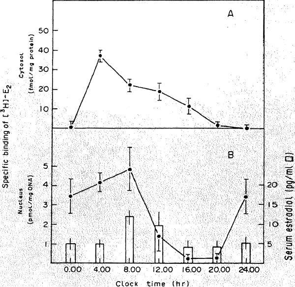
Panel A, Protamine sulfate precipitates of rat liver cytosol were prepared and incubated with varying concentrations of [3H]E2 in the absence (total binding) and presence (nonspecific binding) of a 100-fold excess of unlabeled DES for 16 hr at 4°C. Specific binqing is calculated as the difference of these two values. Each point is the mean ± S.E.M. of cER in five animals, using duplicate determinations for each animal. Scatchard plots of the specific binding values were used to determine the number of binding sites and affinity of binding. Panel B, Nuclear suspensions were incubated with 10 nM [3H]-E2 in the absebce and presence of a 250-fold excess of unlabeled DES for 1 hr at 30°C, as detailed in Methods. Also shown (open bars) are the serum E2 concentrations as determined by radioimmunoassay.
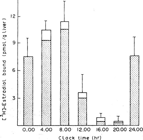
The height of each bar represents the total receptor content (cytosolic and nuclear) corrected for and expressed as binding per gram of liver. The shaded area of each bar represents the contribution of the nuclear receptor to the total amount.
Similar articles
-
Circadian rhythm of hepatic cytosolic and nuclear estrogen and androgen receptors.Gastroenterology. 1986 Jul;91(1):182-8. doi: 10.1016/0016-5085(86)90456-7. Gastroenterology. 1986. PMID: 3710067 Free PMC article.
-
Diurnal variation of cytosol estrogen receptors in the liver from adult male rats.Horm Metab Res. 1983 May;15(5):237-40. doi: 10.1055/s-2007-1018681. Horm Metab Res. 1983. PMID: 6683702
-
Regenerating rat liver: correlations between estrogen receptor localization and deoxyribonucleic acid synthesis.Gastroenterology. 1984 Mar;86(3):552-7. Gastroenterology. 1984. PMID: 6693017 Free PMC article.
-
Estrogen receptor in the mammalian liver.Adv Steroid Biochem Pharmacol. 1979;7:91-117. Adv Steroid Biochem Pharmacol. 1979. PMID: 400323 Review. No abstract available.
-
Estrogen receptors and androgen receptors in the mammalian liver.J Steroid Biochem. 1987;27(4-6):1109-18. doi: 10.1016/0022-4731(87)90197-x. J Steroid Biochem. 1987. PMID: 3320548 Review.
Cited by
-
The Pathophysiologic Role of Disrupted Circadian and Neuroendocrine Rhythms in Breast Carcinogenesis.Endocr Rev. 2016 Oct;37(5):450-466. doi: 10.1210/er.2015-1133. Epub 2016 Jul 26. Endocr Rev. 2016. PMID: 27712099 Free PMC article. Review.
-
Estrogens and the circadian system.Semin Cell Dev Biol. 2022 Jun;126:56-65. doi: 10.1016/j.semcdb.2021.04.010. Epub 2021 May 9. Semin Cell Dev Biol. 2022. PMID: 33975754 Free PMC article. Review.
-
Gastric 17β-estradiol in portal vein and liver Esr1 make a circadian rhythm in systemic circulation in male rats.Endocrine. 2016 Aug;53(2):565-73. doi: 10.1007/s12020-016-0971-0. Epub 2016 May 10. Endocrine. 2016. PMID: 27165169
References
-
- Eagon PK, Porter LE, Francavilla A, DiLeo A, Van Thiel D. Estrogen and androgen receptors in liver: their role in liver disease and regeneration. Semin Liver Dis. 1985;5:59–69. - PubMed
-
- Eagon PK, Fisher SE, Imhoff AF, Porter LE, Stewart RR, Van Thiel DH, Lester R. Estrogen binding proteins of male rat liver: influence of hormonal changes. Arch Biochem Biophys. 1980;201:486–499. - PubMed
-
- Eagon PK, Zdunek JR, Van Thiel DH, Singletary BK, Egler KM, Gavaler JS, Porter LE. Alcohol induced changes in hepatic estrogen binding proteins. Arch Biochem Biophys. 1981;211:48–55. - PubMed
-
- Aten RF, Dickson RB, Eisenfe1d AJ. Estrogen receptors in adult male rat liver. Endocrinology. 1978;103:1629–1635. - PubMed
-
- Powell-Jones W, Thompson C, Nayfeh SN, Lucier GW. Sex differences in estrogen binding by cytosolic and nuclear components of rat liver. J Steroid Biochem. 1980;13:219–229. - PubMed
Publication types
MeSH terms
Substances
Grants and funding
LinkOut - more resources
Full Text Sources