

Methods for Spatiotemporal Analysis of Human Gait Based on Data from Depth Sensors
- PMID: 36772257
- PMCID: PMC9919326
- DOI: 10.3390/s23031218
Methods for Spatiotemporal Analysis of Human Gait Based on Data from Depth Sensors
Abstract
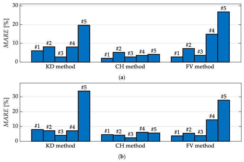
Gait analysis may serve various purposes related to health care, such as the estimation of elderly people's risk of falling. This paper is devoted to gait analysis based on data from depth sensors which are suitable for use both at healthcare facilities and in monitoring systems dedicated to household environments. This paper is focused on the comparison of three methods for spatiotemporal gait analysis based on data from depth sensors, involving the analysis of the movement trajectories of the knees, feet, and centre of mass. The accuracy of the results obtained using those methods was assessed for different depth sensors' viewing angles and different types of subject clothing. Data were collected using a Kinect v2 device. Five people took part in the experiments. Data from a Zebris FDM platform were used as a reference. The obtained results indicate that the viewing angle and the subject's clothing affect the uncertainty of the estimates of spatiotemporal gait parameters, and that the method based on the trajectories of the feet yields the most information, while the method based on the trajectory of the centre of mass is the most robust.
Keywords: data processing; depth sensor; gait analysis; health care; in-home monitoring.
Conflict of interest statement
The authors declare no conflict of interest.
Figures


Similar articles
-
Effects of camera viewing angles on tracking kinematic gait patterns using Azure Kinect, Kinect v2 and Orbbec Astra Pro v2.Gait Posture. 2021 Jun;87:19-26. doi: 10.1016/j.gaitpost.2021.04.005. Epub 2021 Apr 5. Gait Posture. 2021. PMID: 33878509
-
A review of foot pose and trajectory estimation methods using inertial and auxiliary sensors for kinematic gait analysis.Biomed Tech (Berl). 2020 Jun 25:/j/bmte.ahead-of-print/bmt-2019-0163/bmt-2019-0163.xml. doi: 10.1515/bmt-2019-0163. Online ahead of print. Biomed Tech (Berl). 2020. PMID: 32589591 Review.
-
Spatio-temporal gait analysis in children with cerebral palsy using, foot-worn inertial sensors.Gait Posture. 2014;39(1):436-42. doi: 10.1016/j.gaitpost.2013.08.029. Epub 2013 Sep 1. Gait Posture. 2014. PMID: 24044970
-
Improved kinect-based spatiotemporal and kinematic treadmill gait assessment.Gait Posture. 2017 Jan;51:77-83. doi: 10.1016/j.gaitpost.2016.10.001. Epub 2016 Oct 4. Gait Posture. 2017. PMID: 27721202
-
In-Home Older Adults' Activity Pattern Monitoring Using Depth Sensors: A Review.Sensors (Basel). 2022 Nov 23;22(23):9067. doi: 10.3390/s22239067. Sensors (Basel). 2022. PMID: 36501769 Free PMC article. Review.
Cited by
-
Machine Learning-Based Computer Vision for Depth Camera-Based Physiotherapy Movement Assessment: A Systematic Review.Sensors (Basel). 2025 Mar 5;25(5):1586. doi: 10.3390/s25051586. Sensors (Basel). 2025. PMID: 40096440 Free PMC article.
-
Estimation of Reference Values of Gait Spatiotemporal and Kinematic Parameters in the Lower Extremities and Trunk Using a Markerless Motion Capture System for Healthy Older Japanese Adults.Phys Ther Res. 2023;26(3):106-113. doi: 10.1298/ptr.E10247. Epub 2023 Sep 8. Phys Ther Res. 2023. PMID: 38125291 Free PMC article.
-
Minimum required distance for clinically significant measurement of habitual gait speed.BMC Geriatr. 2025 Jul 5;25(1):497. doi: 10.1186/s12877-025-06064-8. BMC Geriatr. 2025. PMID: 40618062 Free PMC article.
-
Automated Gait Analysis Based on a Marker-Free Pose Estimation Model.Sensors (Basel). 2023 Jul 18;23(14):6489. doi: 10.3390/s23146489. Sensors (Basel). 2023. PMID: 37514783 Free PMC article.
References
-
- Montero-Odasso M., Camicioli R., editors. Falls and Cognition in Older Persons: Fundamentals, Assessment and Therapeutic Options. Springer International Publishing; Cham, Switzerland: 2020. Falls as a manifestation of brain failure: Gait, cognition, and the neurobiology of falls; pp. 3–20.
-
- Newman A.B., Simonsick E.M., Naydeck B.L., Boudreau R.M., Kritchevsky S.B., Nevitt M.C., Pahor M., Satterfield S., Brach J.S., Studenski S.A., et al. Association of long-distance corridor walk performance with mortality, cardiovascular disease, mobility limitation, and disability. JAMA. 2006;295:2018–2026. doi: 10.1001/jama.295.17.2018. - DOI - PubMed
-
- Ewins D., Collins T. Clinical Gait Analysis. In: Taktak A., Ganney P., Long D., White P., editors. Clinical Engineering. Academic Press; Oxford, UK: 2014. pp. 389–406.
MeSH terms
LinkOut - more resources
Full Text Sources
Medical
