. 2023 Apr 2:650:81-86.
doi: 10.1016/j.bbrc.2023.02.010. Epub 2023 Feb 6.

HDAC8 regulates protein kinase D phosphorylation in skeletal myoblasts in response to stress signaling

Affiliations

HDAC8 regulates protein kinase D phosphorylation in skeletal myoblasts in response to stress signaling

Justine S Habibian et al. Biochem Biophys Res Commun. .

Abstract

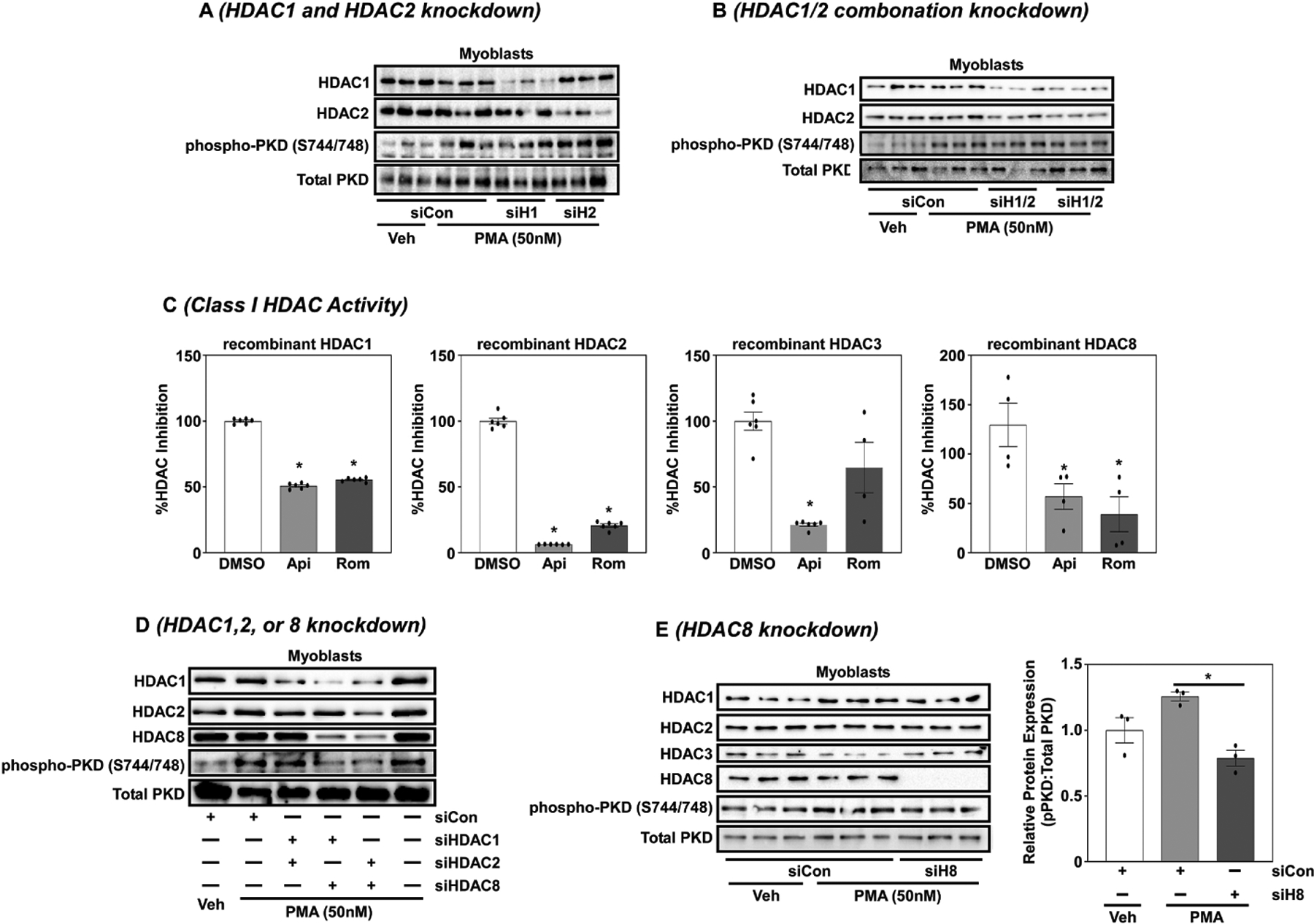
Skeletal muscle differentiation involves activation of quiescent satellite cells to proliferate, differentiate and fuse to form new myofibers; this requires coordination of myogenic transcription factors. Myogenic transcription is tightly regulated by various intracellular signaling pathways, which include members of the protein kinase D (PKD) family. PKD is a family of serine-threonine kinases that regulate gene expression, protein secretion, cell proliferation, differentiation and inflammation. PKD is a unique PKC family member that shares distant sequence homology to calcium-regulated kinases and plays an important role in muscle physiology. In this report, we show that class I histone deacetylase (HDAC) inhibition, and in particular HDAC8 inhibition, attenuated PKD phosphorylation in skeletal C2C12 myoblasts in response to phorbol ester, angiotensin II and dexamethasone signaling independent of changes in total PKD protein expression. As class I HDACs and PKD signaling are requisite for myocyte differentiation, these data suggest that HDAC8 functions as a potential feedback regulator of PKD phosphorylation to control myogenic gene expression.

Keywords: HDAC; HDAC inhibitors; Histone deacetylase; Myoblast; PKD; Protein kinase D.


Conflict of interest statement

Declaration of competing interest The authors declare that they have no known competing financial interests or personal relationships that could have appeared to influence the work reported in this paper.

Figures

Figure 1.
Figure 1.. Class I HDAC inhibition attenuated PKD phosphorylation (S744/S748) in C2C12 myoblasts.
A) C2C12 myoblasts and myotubes (d4) were stimulated with phorbol-12-myristate-13-acetate (PMA; 50nM) and cells lysed over time. Immunoblotting was performed to examine phosphorylated PKD (phospho-PKD; S916; S744/S748) and total PKD protein expression. B). Myoblasts and day 4 differentiated myotubes were pretreated with TSA (200 nM) for 24 hrs prior to stimulation with PMA (50 nM). Cells were lysed and immunoblotted to examine phosphorylated (S916 and S744/S748) and total PKD as well as acetylated histone (acetyl-histone) and acetylated tubulin (acetyl-tubulin). C) Myoblasts were pre-treated with TSA (200nM), apicidin (1uM), DPAH (10uM), Tubastatin A (TubA: 1uM) for 24hrs prior to 30 min PMA (50nM) stimulation. Cells were lysed and immunoblotted for phosphorylated and total PKD (S744/S748), acetyl-histone and acetyl-tubulin. D) Myoblasts were pre-treated with romidepsin (Rom; 1um) or RGFP966 (RGFP; 1uM) for 24hrs prior to 30 min PMA (50nM) stimulation. Cells were lysed and immunoblotted for phosphorylated and total PKD (S744/S748), acetyl-histone and acetyl-tubulin.
Figure 2.
Figure 2.. Class I HDAC inhibition attenuated stress-induced signaling in C2C12 myoblasts.
A) C2C12 myoblasts were stimulated with Dexamethasone (Dex; 1uM) of angiotensin II (AngII; 10uM) and cells lysed over time. Immunoblotting was performed to examine phospho-PKD (S744/S748) and total PKD protein expression and image J used to quantify relative changes in PKD phosphorylation overtime. GraphPad Prism software was used for graph generation. B) Myoblasts were treated with vehicle, Dex (1μM) or AngII (10 μM) for 15 minutes (n=3/treatment) prior to immunoblotting and quantification of phospho-PKD (S744/S748) normalized to total PKD. GraphPad software was used to generate graphs with student’s t-test with Welch’s correction used to asses significance (p<0.05). C) Myoblasts were pretreated with Rom (1uM) for 24hrs prior to stimulation with Dex (1uM) or AngII (10uM). 15 minutes post-treatment cells were lysed and immunoblotted for phospho-PKD (S744/S748) and total PKD protein expression and quantified with Image J software. Graphs were generated and statistical analysis performed via GraphPad software. Student’s t-test with Welch’s correction was used to asses significance (p<0.05).
Figure 3.
Figure 3.. HDAC8 regulates PKD phosphorylation in C2C12 myoblasts in response to PMA signaling.
A) C2C12 myoblasts were transfected with a non-targeting control silencing RNA (siControl) or siRNA targeting HDAC1 (siH1) and HDAC2 (siH2) for 72hrs and then stimulated with PMA (50nM) for 30mins. Cells were lysed and immunoblotted for HDAC1, HDAC2, phosphorylated PKD (phopho-PKD; S744/S748) and total PKD. B) C2C12 myoblasts were transfected with an siControl or two different siRNAs targeting HDACs1 and 2 in combination (sH1/2) for 72hrs prior to PMA (50nM) stimulation for 30min. Cells were lysed and immunoblotted for HDAC1, HDAC2, phospho-PKD (S744/S748) and total PKD. C) Recombinant HDAC1, HDAC2, HDAC3 and HDAC8 were incubated with a class I-selective HDAC substrate prior to fluorescent analysis via the BioTeck Synergy 2 plate reader. D) Myoblasts were transfected with siControl or silencing RNAs in combination of HDACs 1, 2 or 8 for 72hrs. Cells were then stimulated with PMA (50nM) for 30 min, lysed and immunoblotted for HDAC1, HDAC2, HDAC8, phospho-PKD (S744/S748) and total PKD. E) Myoblasts were transfected with siControl or silencing RNA for HDAC8 for 72hrs. Cells were then stimulated with PMA (50nM) for 30min, lysed and immunoblotted for HDAC1, HDAC2, HDAC3, HDAC8, phospho-PKD (S744/S748) and total PKD. Images were quantified using Image J software and graphs were generated and statistical analysis performed via GraphPad software. One-way ANOVA with Tukey’s post-hoc analysis was used to determine significance (p<0.05).

References

    1. Lian D, Chen MM, Wu H, Deng S, Hu X, The Role of Oxidative Stress in Skeletal Muscle Myogenesis and Muscle Disease, Antioxidants. 11 (2022). 10.3390/ANTIOX11040755. - DOI - PMC - PubMed
    1. Seynnes O, Schiaffino S, Blottner D, Csapo R, Gumpenberger M, Wessner B, Skeletal Muscle Extracellular Matrix – What Do We Know About Its Composition, Regulation, and Physiological Roles? A Narrative Review, Front. Physiol 11 (2020) 253. 10.3389/fphys.2020.00253. - DOI - PMC - PubMed
    1. Kleger A, Loebnitz C, Pusapati GV, Armacki M, ller MM, Tümpel S, Illing A, Hartmann D, Brunner C, Liebau S, Rudolph KL, Adler G, Seufferlein T, Protein Kinase D2 Is an Essential Regulator of Murine Myoblast Differentiation, PLoS One. 6 (2011) e14599. 10.1371/JOURNAL.PONE.0014599. - DOI - PMC - PubMed
    1. Yu D, Cai Z, Li D, Zhang Y, He M, Yang Y, Liu D, Xie W, Li Y, Xiao W, Myogenic Differentiation of Stem Cells for Skeletal Muscle Regeneration, Stem Cells Int. 2021 (2021). 10.1155/2021/8884283. - DOI - PMC - PubMed
    1. Czifra G, Tóth IB, Marincsák R, Juhász I, Kovács I, Ács P, Kovács L, Blumberg PM, Bíró T, Insulin-like growth factor-I-coupled mitogenic signaling in primary cultured human skeletal muscle cells and in C2C12 myoblasts. A central role of protein kinase Cδ, (2006). 10.1016/j.cellsig.2005.11.007. - DOI - PubMed

Publication types

MeSH terms