Review
. 2023 Feb 11;14(2):105.
doi: 10.1038/s41419-023-05639-w.

Copper homeostasis and copper-induced cell death in the pathogenesis of cardiovascular disease and therapeutic strategies

Affiliations
Review

Copper homeostasis and copper-induced cell death in the pathogenesis of cardiovascular disease and therapeutic strategies

Xinyue Chen et al. Cell Death Dis. .

Abstract

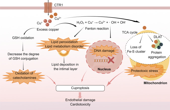
Copper is a vital mineral, and an optimal amount of copper is required to support normal physiologic processes in various systems, including the cardiovascular system. Over the past few decades, copper-induced cell death, named cuproptosis, has become increasingly recognized as an important process mediating the pathogenesis and progression of cardiovascular disease (CVD), including atherosclerosis, stroke, ischemia-reperfusion injury, and heart failure. Therefore, an in-depth understanding of the regulatory mechanisms of cuproptosis in CVD may be useful for improving CVD management. Here, we review the relationship between copper homeostasis and cuproptosis-related pathways in CVD, as well as therapeutic strategies addressing copper-induced cell death in CVD.


Conflict of interest statement

The authors declare no competing interests.

Figures

Fig. 1
Fig. 1. Copper metabolism in mammals at a molecular level.
Cu+ can be sequestered by MT for storage. CTR1 is highly specific for the uptake of Cu+. At physiologic Cu+ levels, copper-transporting ATPases localize in the TGN, where they pump Cu+ from the cytoplasm into the lumen of the TGN. When intracellular Cu+ increases, these copper-transporting ATPases fuse with the plasma membrane to export Cu+. In the basolateral membrane of enterocytes, copper is pumped by ATP7A into the portal circulation and enters the main organ of copper storage, the liver. Excess copper in liver cells is secreted into bile in the form of vesicles via ATP7B. Cu+ travels through the copper transport ATP7B-TGN pathway to form CP, which is then transported to various systems throughout the body. In addition, ATOX1 transports Cu+ to the nucleus, where it binds to transcription factors and drives gene expression. COX17 transports Cu+ to the copper-carrying proteins SCO1, SCO2, and COX11 and delivers it to CCO to activate the activity of enzymes in the respiratory chain. CCS can transfer Cu+ to SOD1. ATOX1 antioxidant-1, ATP7A copper-transporting ATPase alpha, ATP7B copper-transporting ATPase beta, CCO cytochrome C oxidase, CTR1 copper transporter 1 of CCO, CCS Cu chaperone for SOD1, COX11 cytochrome c oxidase copper chaperone 11, COX17 cytochrome C oxidase copper chaperone 17, CP ceruloplasmin, GSH glutathione, MT metallothionein, SCO1 synthesis of cytochrome C oxidase 1, SCO2 synthesis of cytochrome C oxidase 2, SOD1 superoxide dismutase 1, TGN trans Golgi network. The figure was created with Figdraw (https://www.figdraw.com/).
Fig. 2
Fig. 2. Oxidative stress induced by copper-induced cell death in CVD.
Excess copper results in the oxidation of catecholamines by promoting GSH oxidation. Through the Fenton reaction, copper produces oxidative stress, increasing lipid metabolism dysfunction and leading to DNA breakage. Copper ions directly bind fatty acylation components in the TCA cycle, leading to the aggregation and dysregulation of these proteins, blocking the TCA cycle of the tricarboxylic acid cycle, triggering proteotoxic stress, and inducing cell death. The above mechanisms may lead to endothelial injury and cardiotoxicity. CVD cardiovascular disease, CTR1 calcitonin receptor 1, DLAT dihydrolipoamide S-acetyltransferase, Fe-S iron-sulfur proteins, GSH glutathione, S sulfur ion, TCA tricarboxylic acid cycle. The figure was created with Figdraw (https://www.figdraw.com/).
Fig. 3
Fig. 3. Mitochondria and copper in CVD.
When copper deficiency reduces the activity of CCO, the level of ATP and phosphocreatine in the heart and other tissues decreases, whereas the content of ADP, orthophosphate, glycogen, and lipid droplets increases. Mitochondrial cristae and inner and outer membranes are altered, eventually leading to mitochondrial rupture. Additionally, copper deficiency increases the level of PGC-1α, causing mitochondrial dysfunction. These changes interfere with energy metabolism and cause myocardial damage. CVD cardiovascular disease, CCO cytochrome C oxidase, COX11 cytochrome c oxidase copper chaperone 11, COX17 cytochrome C oxidase copper chaperone 17, PGC-1α peroxisome proliferator-activated receptor-gamma coactivator-1 alpha protein, SCO1 synthesis of cytochrome C oxidase 1, SCO2 synthesis of cytochrome C oxidase 2. The figure was created with Figdraw (https://www.figdraw.com/).
Fig. 4
Fig. 4. Vascular regulation and copper in CVD.
Copper can regulate the activity of HIF-1. HIF-1 consists of HIF-1α and HIF-1β. CCS transports copper into the nucleus, and CuBP mediates subsequent actions of copper. The core base “GGAA” (the core motif of the ETS family) is a key motif in the binding site of copper-dependent genes. p300, CBP, and SRC1 act as cofactors to form the HIF-1 transcription complex. The interaction of HIF-1 with HRE requires copper to initiate the copper-dependent expression of genes such as VEGF. LOX is essential for vascular maturation. Copper can regulate LOX production through ATOX1, ATP7A, and RAC1. Ischemia and hypoxia increase the efflux of copper. The inhibition of the above mechanisms caused by copper efflux will bring about vascular wall hypotonia, increased myocardial fragility, and angiogenesis depression, and will eventually lead to myocardial damage. ATOX1 antioxidant 1 copper chaperone, ATP7A ATPase copper transporting alpha, BNIP3 BCL2 interacting protein 3, CCS Cu chaperone for SOD1, CTR1 calcitonin receptor 1, ETS E-twenty-six, HIF hypoxia inducible factor, HRE hypoxia-responsive element, LOX lysyl oxidase, RAC1 ras-related C3 botulinum toxin substrate 1, TGN trans Golgi network, VEGF vascular endothelial growth factor. The figure was created with Figdraw (https://www.figdraw.com/).

References

    1. Kesavardhana S, Malireddi RKS, Kanneganti TD. Caspases in cell death, inflammation, and pyroptosis. Annu Rev Immunol. 2020;38:567–95. doi: 10.1146/annurev-immunol-073119-095439. - DOI - PMC - PubMed
    1. Chen L, Min J, Wang F. Copper homeostasis and cuproptosis in health and disease. Signal Transduct Target Ther. 2022;7:378. doi: 10.1038/s41392-022-01229-y. - DOI - PMC - PubMed
    1. Wang N, Xu X, Li H, Feng Q, Wang H, Kang YJ. Atherosclerotic lesion-specific copper delivery suppresses atherosclerosis in high-cholesterol-fed rabbits. Exp Biol Med. 2021;246:2671–8. doi: 10.1177/15353702211046541. - DOI - PMC - PubMed
    1. Zhang J, Cao J, Zhang H, Jiang C, Lin T, Zhou Z, et al. Plasma copper and the risk of first stroke in hypertensive patients: a nested case-control study. Am J Clin Nutr. 2019;110:212–20. doi: 10.1093/ajcn/nqz099. - DOI - PubMed
    1. Powell SR, Hall D, Shih A. Copper loading of hearts increases postischemic reperfusion injury. Circ Res. 1991;69:881–5. doi: 10.1161/01.RES.69.3.881. - DOI - PubMed

Publication types