. 2023 Jun;242(6):1012-1028.
doi: 10.1111/joa.13831. Epub 2023 Feb 11.

Three-dimensional correlative light and focused ion beam scanning electron microscopy reveals the distribution and ultrastructure of lanceolate nerve endings surrounding terminal hair follicles in human scalp skin

Affiliations

Three-dimensional correlative light and focused ion beam scanning electron microscopy reveals the distribution and ultrastructure of lanceolate nerve endings surrounding terminal hair follicles in human scalp skin

Haruyo Yamanishi et al. J Anat. 2023 Jun.

Abstract

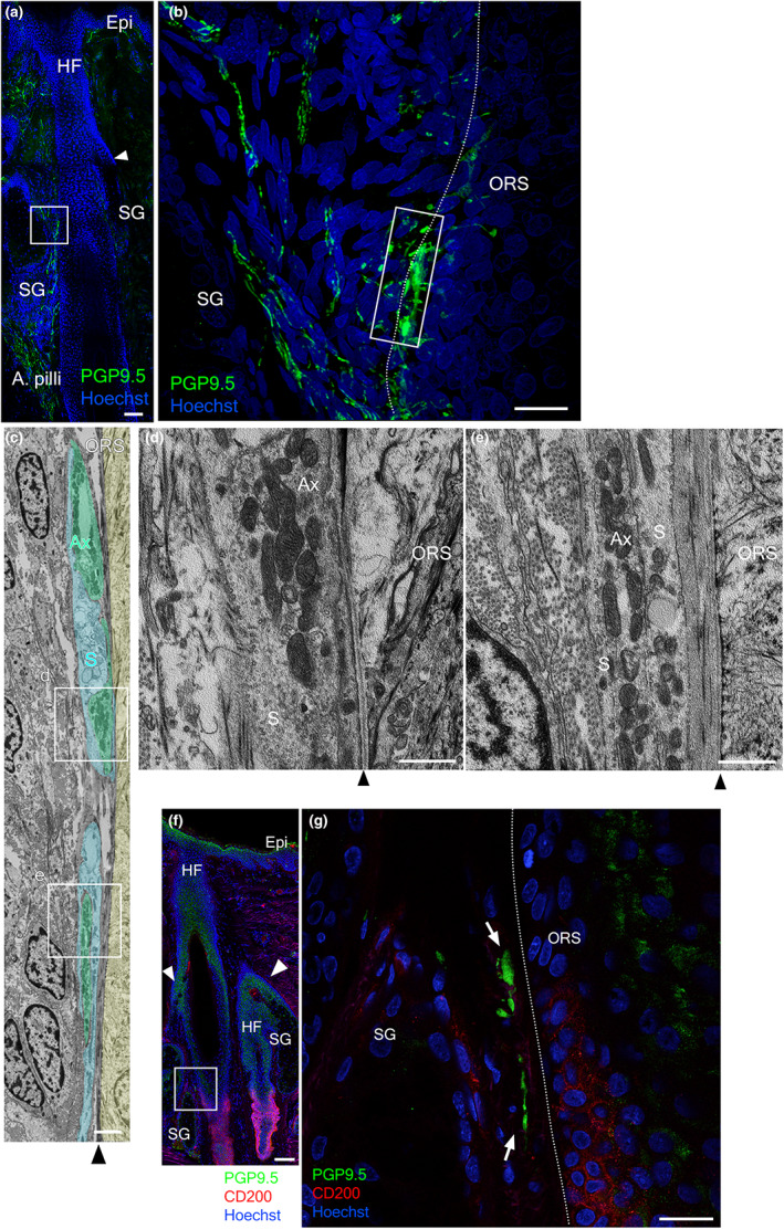
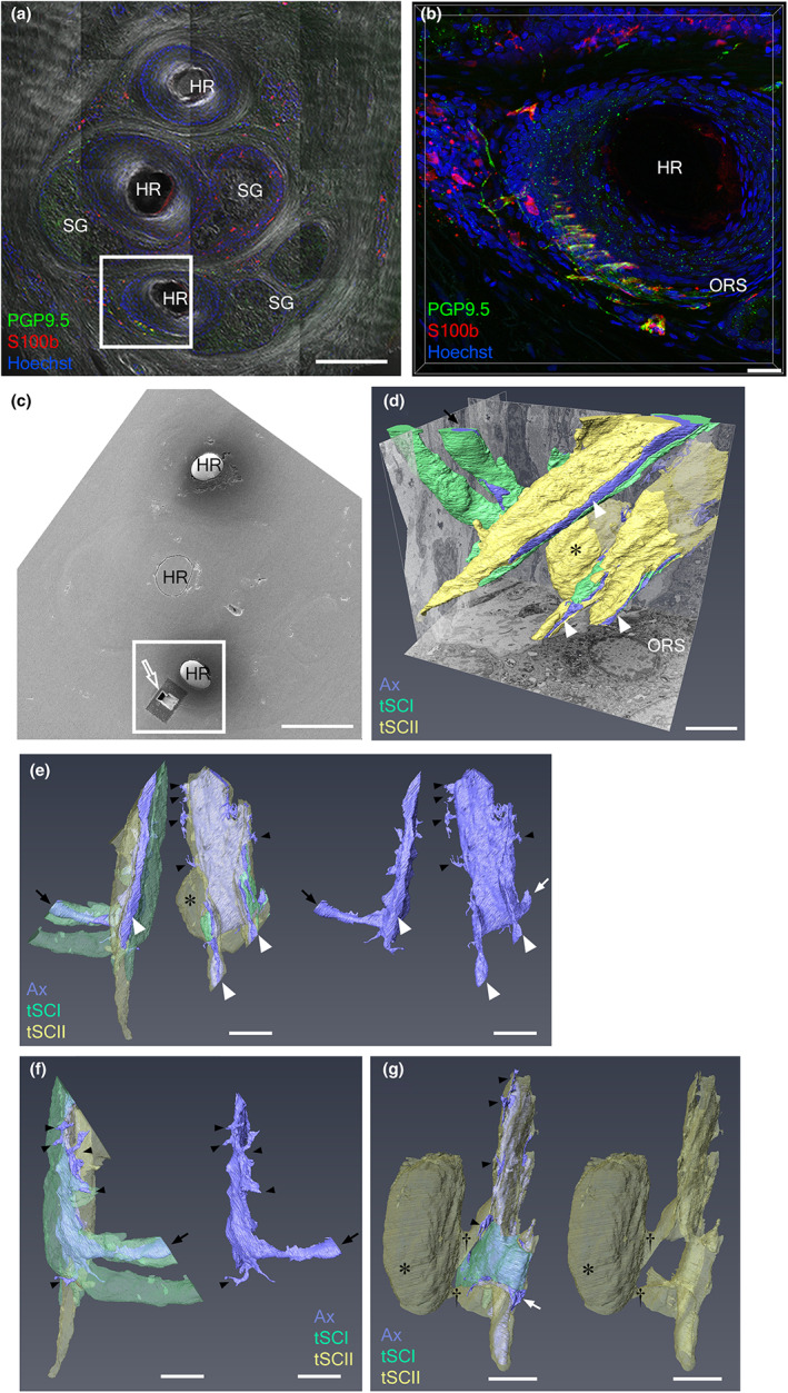
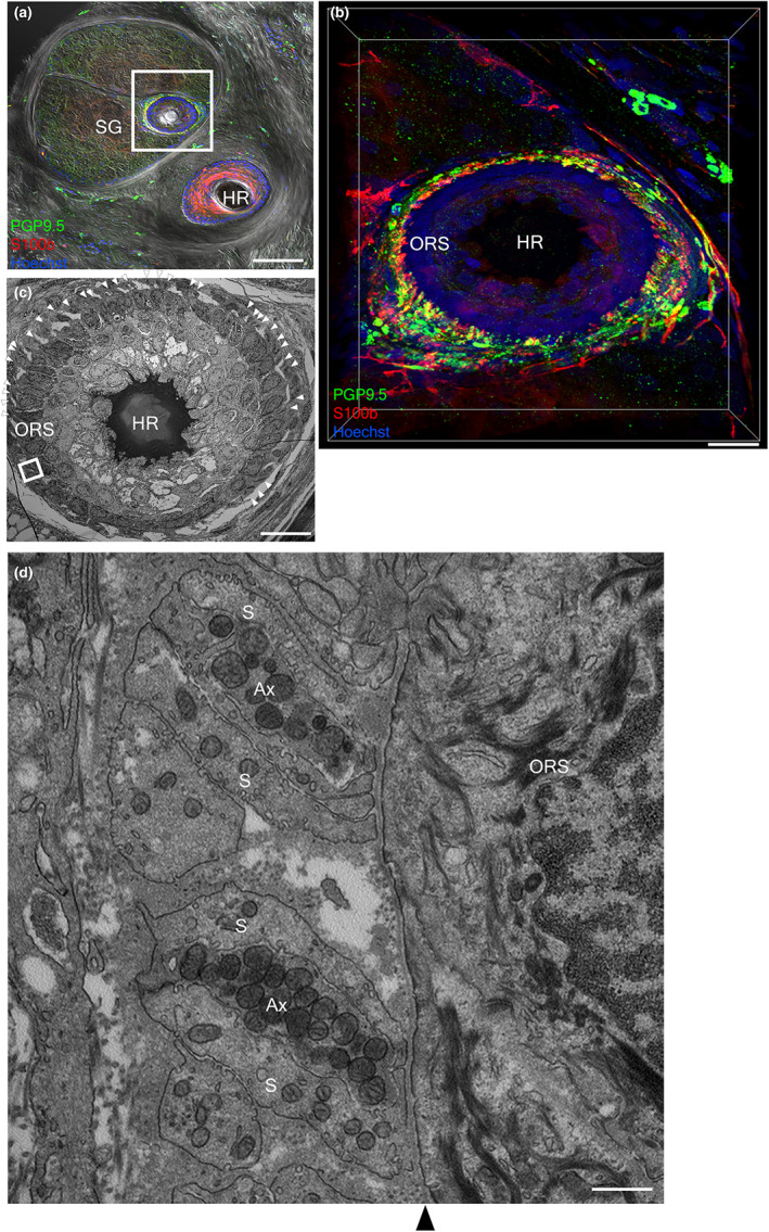
Lanceolate nerve endings (LNEs) surrounding hair follicles (HFs) play an important role in detecting hair deflection. Complexes of the LNEs form a palisade-like structure along the longitudinal axis of hair roots in which axons are sandwiched between two processes of terminal Schwann cells (tSCs) at the isthmus of HFs. The structure and molecular mechanism of LNEs in animal sinus hair, pelage, and human vellus hairs have been investigated. Despite the high density of HFs in human scalp skin, the LNEs in human terminal HFs have not been investigated. In this study, we aimed to reveal the distribution and ultrastructure of LNEs in terminal HFs of human scalp skin. Using light-sheet microscopy and immunostaining, the LNEs were observed at one terminal HF but not at the other terminal HFs in the same follicular unit. The ultrastructure of the LNEs of terminal HFs in human scalp skin was characterized using correlated light and electron microscopy (CLEM). Confocal laser microscopy and transmission electron microscopy of serial transverse sections of HFs revealed that LNEs were aligned adjacent to the basal lamina outside the outer root sheath (ORS), at the isthmus of terminal HFs, and adjacent to CD200-positive ORS cells in the upper bulge region. Moreover, axons with abundant mitochondria were sandwiched between tSCs. Three-dimensional CLEM, specifically confocal laser microscopy and focused ion beam scanning electron microscopy, of stained serial transverse sections revealed that LNEs were wrapped with type I and type II tSCs, with the processes protruding from the space between the Schwann cells. Moreover, the ultrastructures of LNEs at miniaturized HFs were similar to those of LNEs at terminal HFs. Preembedding immunoelectron microscopy revealed that Piezo-type mechanosensitive ion channel component 2 (Piezo2), a gated ion channel, was in axons and tSCs and adjacent to the cell membrane of axons and tSCs, suggesting that LNEs function as mechanosensors. The number of LNEs increased as the diameter of the ORS decreased, suggesting that LNEs dynamically adapt to the HF environment as terminal HFs miniaturize into vellus-like hair. These findings will provide insights for investigations of mechanosensory organs, aging, and re-innervation during wound healing.

Keywords: correlative light and electron microscopy; focused ion beam scanning electron microscopy; immunoelectron microscopy; lanceolate nerve endings; light-sheet microscopy; miniaturized hair follicles; palisade nerve endings; terminal hair follicles.


Conflict of interest statement

The authros declared that they have no conflict of interest.

Figures

FIGURE 1
FIGURE 1
Schematic illustrations of hair follicles in human scalp skin. (a) A hair follicular unit composed of normal terminal HFs. (b) A transverse section at the isthmus level is indicated by the brown rectangle in panel a. (c) A magnified image of LNEs, which are composed of axons and tSCs. (d) A hair follicular unit including a miniaturized HF. (e) A transverse section at the isthmus level of the miniaturized HF by the brown rectangle in panel d. A pili: arrector pili muscle; HR: hair root; LNEs: lanceolate nerve endings; ORS: outer root sheath; SG: sebaceous gland.
FIGURE 2
FIGURE 2
Neural network at a human scalp hair follicular unit identified by light‐sheet microscopy. (a–d) Whole‐mount immunolabeling with PGP9.5 (green) and S100β (magenta) shows innervation, indicated by PI (red), surrounding HF outside the ORS. (a, b) Neural structures (white arrows) at a terminal hair within a three‐hair follicular unit. (c, d) Palisade‐like lanceolate complex (white bracket) at a miniaturized hair within a three‐hair follicular unit. (b', d') Magnified views of neural structures and LNEs indicated by white rectangles in panels b and d, respectively. A pili: arrector pili muscle; Epi: epidermis; HF: hair follicle; HR: hair root; ORS: out root sheath; SG: sebaceous gland; SwG: sweat gland. Bars = 200 μm (a, b, c, d) and 50 μm (b', d'). Images of scalp skin taken from a 51‐year‐old man.
FIGURE 3
FIGURE 3
LNEs at the terminal HFs by correlative confocal laser and electron microscopy. (a) A transverse section of a two‐hair follicular unit immunolabeled with PGP9.5 (green), S100β (red), and Hoechst (blue) merged with a differential interference contrast image. (b, c) Magnified views of a white rectangle in panel a. LNEs encircle part of an HF. Double staining with PGP9.5 and S100β indicates that LNEs (white arrowheads in b) are adjacent to ORS. (d) The transmission electron micrograph shows the ultrastructure of LNEs in panels a and b. (e) A magnified image of a white rectangle in panel d shows that lanceolate nerve endings are aligned along the basal lamina (black arrowhead) outside the ORS, and axons with abundant mitochondria are sandwiched by terminal Schwann cells. Ax: axon; C, collagen; E: elastin; HR: hair root; ORS: outer root sheath; S: terminal Schwan cells; SG: sebaceous gland. Bars = 200 μm (a), 20 μm (b–d), and 1 μm (e). Images of scalp skin taken from a 51‐year‐old man.
FIGURE 4
FIGURE 4
Cross‐sectioned LNEs at a terminal hair by correlative confocal laser and electron microscopy. (a) Cross‐sectioned terminal HF immunolabeled with PGP9.5 (green) and Hoechst (blue). (b) Magnified view of a rectangle in panel a. (c) Transmission electron micrograph of the white rectangle of panel b. (d, e) Magnified images of white rectangles of panel c. (f) Terminal HF immunolabeled with PGP9.5 (green), CD200 (red), and Hoechst (blue). (g) Magnified view of panel f. A pili: arrector pili muscle; Ax: axon; Epi: epidermis; HF: hair follicle; ORS: outer root sheath; S: terminal Schwan cells; SG: sebaceous gland; Bars = 100 μm (a, f), 20 μm (b, g), 2 μm (c), and 1 μm (d, e). Images of scalp skin taken from a 51‐year‐old man (a–e) and a 38‐year‐old man (f, g).
FIGURE 5
FIGURE 5
Three‐dimensional reconstructed images of LNEs in a terminal HF by 3D‐CLEM. (a) The transverse section of a follicular unit immunolabeled with PGP9.5 (green), S100β (red), and Hoechst (blue) merged with a differential interference contrast image. (b) Magnified view of the white rectangle in panel a. (c) Surface of the three‐hair follicular unit embedded in resin. (d–g) 3D reconstruction of two LNEs marked by the white opened arrow in panel c. Axons (purple) face the basal lamina (white arrowheads). Type I and type II tSCs are marked by green and yellow, respectively. Asterisk marks the nuclei of tSC (d, e, g), and the two processes originating from tSC are marked by daggers (g). Ax: axon; HR: hair root; ORS: outer root sheath; tSC: terminal Schwan cells; SG: sebaceous gland. Bars = 200 μm (a, c), 30 μm (b), 4 μm (e, f, g). Images of scalp skin taken from a 51‐year‐old man.
FIGURE 6
FIGURE 6
LNEs at a miniaturized HF by correlative confocal laser and electron microscopy. (a) The transverse section of a two‐hair follicular unit immunolabeled with PGP9.5 (green), S100β (red), and Hoechst (blue) merged with a differential interference contrast image. (b) Magnified views of the white rectangle in panel a. LNEs encircle the HF. (c) The transmission electron micrograph shows the distribution of LNEs (white arrowheads) in panel b. (d) A magnified image of the white rectangle in panel c shows that LNEs are aligned along the basal lamina (black arrowhead) outside the ORS and sandwiched by tSC. Ax: axon; C: collagen; E: elastin; HR: hair root; ORS: outer root sheath; S: terminal Schwan cells; SG: sebaceous gland. Bars = 100 μm (a), 20 μm (b, c), and 500 nm (d). Images of scalp skin taken from a 48‐year‐old man.
FIGURE 7
FIGURE 7
Cross‐sectioned LNEs in a miniaturized hair follicle by correlative confocal laser and electron microscopy. (a, a') A miniaturized HF immunolabeled with PGP9.5 (green) and Hoechst (blue) in serial sections. (b) A magnified view of a white rectangle in panel a'. (c) Transmission electron micrographs of the white rectangle of panel a'. Axon (green), terminal Schwann cell (pale blue), and ORS (yellow) are assigned pseudo‐colors. (d, e) Magnified images of white rectangles of panel c. Small processes (white open arrows) protrude toward the connective tissue (d). Ax: axon; DP: dermal papillae HF: hair follicle; ORS: outer root sheath; S: terminal Schwan cells; SG: sebaceous gland; white arrowhead: sebaceous duct; black arrowheads: basal lamina. Bars = 100 μm (a), 2 μm (b, c), and 500 nm (d, e). Images of scalp skin taken from a 59‐year‐old man.
FIGURE 8
FIGURE 8
Three‐dimensional reconstructed images of LNEs in a miniaturized hair follicle by 3D‐CLEM. (a) The transverse section of a three‐hair follicular unit immunolabeled with PGP9.5 (green) and Hoechst (blue) merged with a differential interference contrast image. (b) Magnified view of the white dashed rectangle in panel a. White arrows mark LNEs. (c) The surface of the follicular unit embedded in resin indicated by the dashed line in panel a. (d, e) 3D reconstruction of two LNEs in the black rectangle of panel c. Axons (purple) face the basal lamina (black arrows). Nerve endings with branched processes (black arrowheads) are enclosed within terminal Schwann cells (yellow). Ax: axon; HR: hair root; ORS: outer root sheath; tSCII: type II terminal Schwan cells; SG: sebaceous gland. Bars = 200 μm (a), 30 μm (b), 100 μm (c), and 6 μm (d, e). Images of scalp skin taken from a 51‐year‐old man.
FIGURE 9
FIGURE 9
Piezo2 expressed at LNEs and tSCs in the terminal and miniaturized HFs are revealed by preembedding immunoelectron microscopy. (a) The transverse section of a three‐hair follicular unit immunolabeled with Piezo2 (green), PGP9.5 (red), and Hoechst (blue) merged with a differential interference contrast image. (b) Magnified view of the white rectangle in panel a. LNEs (white arrowheads) are located along part of the terminal HF. (c) Distribution of Piezo2 (black arrows) in axons, tSCs, and the lanceolate endings of the terminal HF by preembedding immunoelectron microscopy. (d) The transverse section of a two‐hair follicular unit immunolabeled with Piezo2 (green), PGP9.5 (red), and Hoechst (blue) merged with a differential interference contrast image. (e) Magnified view of the white rectangle in panel a. LNEs (white arrowheads) partially encircle the miniaturized HF. (f) Distribution of Piezo2 (black arrows) in the lanceolate endings of the miniaturized HF by preembedding immunoelectron microscopy. Piezo2 expressed adjacent to the cell membrane is indicated by white arrows (c, f). Ax: axon; HR: hair root; ORS: outer root sheath; S: terminal Schwan cells; SG: sebaceous gland; Asterisk: miniaturized HF. Bars = 100 μm (a, d), 30 μm (b, d), and 500 nm (c, f). Images of scalp skin taken from a 29‐year‐old man.
FIGURE 10
FIGURE 10
Comparison of lanceolate nerve ending morphology between terminal and miniaturized HFs. (a) The transverse section of a two‐hair follicular unit immunolabeled with PGP9.5 (green), S100β (red), and Hoechst (blue) merged with a differential interference contrast image. (b) Distribution of LNEs (white arrowheads) in terminal HFs. (c) The transverse section of a four‐hair follicular unit immunolabeled with PGP9.5 (green), S100β (red), and Hoechst (blue) merged with a differential interference contrast image. (d) Distribution of LNEs (white arrowheads) in a miniaturized HF. (e) Quantitative analysis of the relationship between the number of LNEs and the diameter of ORS in HFs from six donors. HR: hair root; ORS: outer root sheath; SG: sebaceous gland; Asterisk: miniaturized HFs. Bars = 100 μm (a, c) and 20 μm (b, d). Images of scalp skin taken from a 48‐year‐old man.

Similar articles

Cited by

References

    1. Blanpain, C. , Lowry, W.E. , Geoghegan, A. , Polak, L. & Fuchs, E. (2004) Self‐renewal, multipotency, and the existence of two cell populations within an epithelial stem cell niche. Cell, 118, 635–648. - PubMed
    1. Blume, U. , Verschoore, M. , Poncet, M. , Czernielewski, J. , Orfanos, C.E. & Schaefer, H. (1993) The vellus hair follicle in acne: hair growth and sebum excretion. British Journal of Dermatology, 129, 23–27. - PubMed
    1. Cheng, C.C. , Tsutsui, K. , Taguchi, T. , Sanzen, N. , Nakagawa, A. , Kakiguchi, K. et al. (2018) Hair follicle epidermal stem cells define a niche for tactile sensation. eLife, 7, e38883. - PMC - PubMed
    1. Furuta, T. , Bush, N.E. , Yang, A.E. , Ebara, S. , Miyazaki, N. , Murata, K. et al. (2020) The cellular and mechanical basis for response characteristics of identified primary afferents in the rat vibrissal system. Current Biology, 30, 815–826. - PMC - PubMed
    1. Ghitani, N. , Barik, A. , Szczot, M. , Thompson, J.H. , Li, C. , le Pichon, C.E. et al. (2017) Specialized mechanosensory nociceptors mediating rapid responses to hair pull. Neuron, 95, 944–954. - PMC - PubMed