. 2023 Feb 11;15(1):10.
doi: 10.1186/s13089-023-00310-z.

Alveolar capillary dysplasia with misalignment of pulmonary veins in a premature newborn: the role of lung ultrasound

Affiliations

Alveolar capillary dysplasia with misalignment of pulmonary veins in a premature newborn: the role of lung ultrasound

Macarena L Atun et al. Ultrasound J. .

Abstract

Background: Alveolar capillary dysplasia with misalignment of pulmonary veins (ACD/MPV) is a lethal neonatal lung disorder characterized by the decrease of the alveolar units, abnormalities in the air-blood barrier of the lung, and impaired gas exchange. Typically, it affects a full-term newborn; the symptoms usually start within a few hours after birth, resulting in severe respiratory distress and pulmonary hypertension. In most of the cases, this disorder is refractory to conventional pulmonary support.

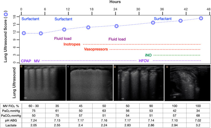
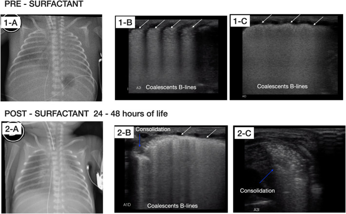
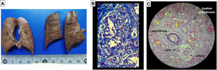
Case presentation: We report a case of a newborn male of 29 weeks gestational age, with birth weight of 850 g and intrauterine growth restriction. Severe respiratory distress appeared a few minutes after birth; non-invasive ventilatory support was provided in the delivery room and, as a consequence of persistent respiratory failure, he was admitted to the neonatal intensive care unit (NICU) where mechanical ventilation was required. Due to the symptoms and pulmonary ultrasound pattern suggestive of respiratory distress syndrome, surfactant treatment was administered. Lung ultrasound (LU) was used for monitoring the responsiveness to surfactant; severe pulmonary hypertension ensued, followed by respiratory failure, refractory shock, and death within 48 h. Owing to the poor response to the established therapy, ACD/MPV was suspected. The diagnosis was confirmed through autopsy. The main goal of this case report is to show the role of LU for monitoring the evolution of this disorder.

Conclusion: LU could provide essential information to help diagnose and follow-up the underlying cause of persistent pulmonary hypertension of the newborn in an earlier and more effective way than chest X-ray. LU is suitable for routine monitoring of lung disease in the NICU.

Keywords: Congenital alveolar dysplasia; Lung ultrasound; Neonate; Pulmonary hypertension; Respiratory distress.


Conflict of interest statement

The authors declare that they have no competing interests.

Figures

Fig. 1
Fig. 1
Medical therapy, LU score trend and clinical parameters during the first 48 h of life. LU pattern representatives for each time. ABGs arterial blood gases, iNO nitric oxide, CPAP continuous positive airway pressure, MV mechanical ventilation, HFOV high-frequency oscillation ventilation
Fig. 2
Fig. 2
Radiographic and LU examination pre-surfactant administration and after 24–48 h of life. Top: chest X-ray and LU examination after birth pre-surfactant administration. 1-A Chest X-ray shows poorly inflated lungs with ground glass shadowing widespread in both hemithorax with obliteration of cardiac silhouette. 1-B and 1-C LU examinations in longitudinal and oblique view, respectively, show irregularity of the pleural line (white arrows), subpleural consolidation and widespread appearance of “white lung” on both lung fields without spared areas. Bottom: chest X-ray and LU examinations performed after 24–48 h of life. 2-A chest X-ray shows increased interstitial infiltrate. 2-B and 2-C LU examinations in oblique view, show severe deterioration of lung aeration, presence of multiple bilateral consolidations (blue arrows), air bronchograms, and coalescent B-lines without spared areas
Fig. 3
Fig. 3
Macroscopic and histopathological finding of alveolar capillary dysplasia with ACD/MPV. A Macroscopic image and the cut surface of the lungs. Histopathology study: B abnormal capillary pattern (red arrows) in the pulmonary interstitial space (Masson’s trichrome stain); C thickening of the alveolar wall (yellow arrows), irregular capillary vascularization, hypertrophy of the medial layer of the arterioles, abnormal arrangement of the veins adjacent to the arteries and foci of hyaline membranes (hematoxylin and eosin stain)

References

    1. Bishop NB, Stankiewicz P, Steinhorn RH. Alveolar capillary dysplasia. Am J Respir Crit Care Med. 2011;184(2):172–179. doi: 10.1164/rccm.201010-1697CI. - DOI - PMC - PubMed
    1. Langston C. Misalignment of pulmonary vein and alveolar capillary dysplasia. Pediatr Pathol. 1991;11:163–170. doi: 10.3109/15513819109064753. - DOI - PubMed
    1. Michalsky MP, Arca MJ, Groenman F, Hammond S, Tibboel D, Caniano DA. Alveolar capillary dysplasia: a logical approach to a fatal disease. J Pediatr Surg. 2005;40:1100–1105. doi: 10.1016/j.jpedsurg.2005.03.067. - DOI - PubMed
    1. Brat R, Yousef N, Klifa R, Reynaud S, Shankar Aguilera S, De Luca D. Lung ultrasonography score to evaluate oxygenation and surfactant need in neonates treated with continuous positive airway pressure. JAMA Pediatr. 2015;169(8):e151797. doi: 10.1001/jamapediatrics.2015.1797. - DOI - PubMed
    1. Slot E, Edel G, Cutz E, Van Heijst A, Post M, Schnater M, Wijnen R, Tibboel D, Rottier R, Klein A. Alveolar capillary dysplasia with misalignment of the pulmonary veins: clinical, histological, and genetic aspects. Pulm Circ. 2018;8(3):1–8. doi: 10.1177/2045894018795143. - DOI - PMC - PubMed

LinkOut - more resources