

The effect of interlaminar Coflex stabilization in the topping-off procedure on local and global spinal sagittal alignment
- PMID: 36774472
- PMCID: PMC9921634
- DOI: 10.1186/s12891-023-06231-1
The effect of interlaminar Coflex stabilization in the topping-off procedure on local and global spinal sagittal alignment
Abstract
Purpose: To investigate the effect of interlaminar Coflex stabilization (ICS) at various segments in the topping-off procedure on local and global spinal sagittal alignment.
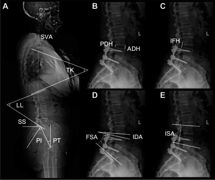
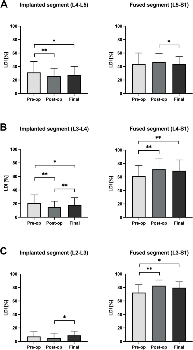
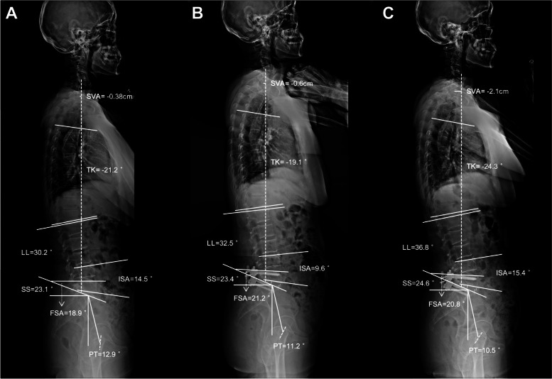
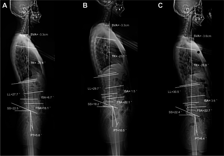
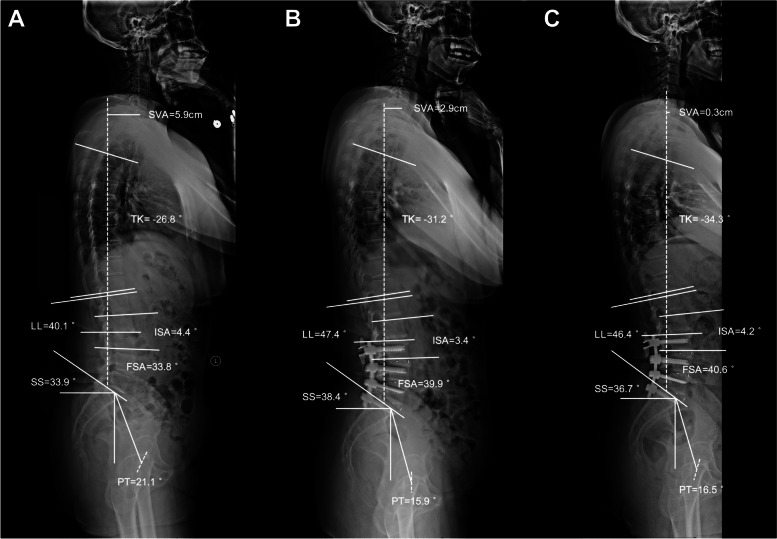
Methods: Eighty-nine consecutive patients with degenerative lumbar spinal stenosis (DLSS) who underwent ICS and transforaminal lumbar interbody fusion (TLIF) were retrospectively reviewed. They were divided into Group A (L4-L5 ICS + L5-S1 TLIF), Group B (L3-L4 ICS + L4-S1 TLIF), and Group C (L2-L3 ICS + L3-S1 TLIF) according to their fusion levels. The measured local sagittal parameters included the implanted segmental angle (ISA), intervertebral disc angle (IDA), intervertebral foreman height (IFH), and disc height. The assessed global sagittal parameters included thoracic kyphosis, lumbar lordosis (LL), the fused segment angle (FSA), the sacral slope, the pelvic tilt, pelvic incidence, and the sagittal vertical axis. The Oswestry Disability Index (ODI) and visual analog scales (VAS) were recorded to evaluate the clinical outcomes.
Results: Regarding the local alignment parameters, the ISA and IDA decreased immediately after surgery in Groups A and B, followed by an increase at the last follow-up (all, P < 0.05). Conversely, the IFH of Groups A and B first increased after surgery and then decreased to approximately the original value (all, P < 0.05). No significant differences were evident between the local sagittal parameters at different time points in Group C. Regarding the global sagittal profiles, the LL and FSA exhibited a significant postoperative increase (both at P < 0.05) in all the groups. All three groups displayed significant improvements in the ODI, VAS-back pain, and VAS-leg pain. Furthermore, 4.5% (4/89) of the patients exhibited radiographic adjacent segment degeneration (ASD) at the last follow-up.
Conclusion: ICS during topping-off surgery led to a temporary loss of local lordosis, especially in the lower lumbar segment, while the intervertebral space realigned after middle-term follow-up. The topping-off procedure with ICS is a feasible and promising surgical option of DLSS since it reduces fusion levels and prevents ASD development.
Keywords: Coflex; Degenerative lumbar spinal stenosis; Interlaminar dynamic stabilization; Sagittal spinal alignment; Topping-off procedure.
© 2023. The Author(s).
Conflict of interest statement
The authors declare no competing interests.
Figures
Similar articles
-
Topping-off surgery vs posterior lumbar interbody fusion for degenerative lumbar disease: a comparative study of clinical efficacy and adjacent segment degeneration.J Orthop Surg Res. 2019 Jun 28;14(1):197. doi: 10.1186/s13018-019-1245-3. J Orthop Surg Res. 2019. PMID: 31253158 Free PMC article.
-
Radiological adjacent-segment degeneration in L4-5 spondylolisthesis: comparison between dynamic stabilization and minimally invasive transforaminal lumbar interbody fusion.J Neurosurg Spine. 2018 Sep;29(3):250-258. doi: 10.3171/2018.1.SPINE17993. Epub 2018 Jun 1. J Neurosurg Spine. 2018. PMID: 29856306
-
How to prevent preoperative adjacent segment degeneration L5/S1 segment occuring postoperative adjacent segment disease? A retrospective study of risk factor analysis.J Orthop Surg Res. 2025 Mar 10;20(1):259. doi: 10.1186/s13018-024-05439-8. J Orthop Surg Res. 2025. PMID: 40065447 Free PMC article. Review.
-
Comparative analysis of the effects of OLIF and TLIF on adjacent segments after treatment of L4 degenerative lumbar spondylolisthesis.J Orthop Surg Res. 2022 Apr 4;17(1):203. doi: 10.1186/s13018-022-03084-7. J Orthop Surg Res. 2022. PMID: 35379259 Free PMC article.
-
Degenerative lumbar spondylolisthesis: cohort of 670 patients, and proposal of a new classification.Orthop Traumatol Surg Res. 2014 Oct;100(6 Suppl):S311-5. doi: 10.1016/j.otsr.2014.07.006. Epub 2014 Sep 5. Orthop Traumatol Surg Res. 2014. PMID: 25201282 Review.
Cited by
-
Correlation between neurogenic injury and lumbar disc degeneration in tethered cord syndrome: a retrospective MRI study based on somatosensory evoked potentials.Eur Spine J. 2025 Sep 5. doi: 10.1007/s00586-025-09339-1. Online ahead of print. Eur Spine J. 2025. PMID: 40911075
-
The clinical efficacy of hybrid surgery based on the Waveflex semi-rigid dynamic internal fixation system for the treatment of lumbar degenerative diseases: over three-year follow-up study.BMC Musculoskelet Disord. 2025 May 10;26(1):463. doi: 10.1186/s12891-025-08715-8. BMC Musculoskelet Disord. 2025. PMID: 40349030 Free PMC article.
-
The biomechanical changes of facet joint violation after transforaminal lumbar interbody fusion combined with decompression surgery: a finite element study.Front Bioeng Biotechnol. 2025 Aug 1;13:1481719. doi: 10.3389/fbioe.2025.1481719. eCollection 2025. Front Bioeng Biotechnol. 2025. PMID: 40821663 Free PMC article.
References
-
- Okuda S, Nagamoto Y, Matsumoto T, Sugiura T, Takahashi Y, Iwasaki M. Adjacent Segment Disease After Single Segment Posterior Lumbar Interbody Fusion for Degenerative Spondylolisthesis: Minimum 10 Years Follow-up. Spine (Phila Pa 1976) 2018;43(23):E1384–e1388. doi: 10.1097/BRS.0000000000002710. - DOI - PubMed
MeSH terms
Grants and funding
LinkOut - more resources
Full Text Sources
