

Spermatogenesis associated serine rich 2 like plays a prognostic factor and therapeutic target in acute myeloid leukemia by regulating the JAK2/STAT3/STAT5 axis
- PMID: 36774517
- PMCID: PMC9921581
- DOI: 10.1186/s12967-023-03968-0
Spermatogenesis associated serine rich 2 like plays a prognostic factor and therapeutic target in acute myeloid leukemia by regulating the JAK2/STAT3/STAT5 axis
Abstract
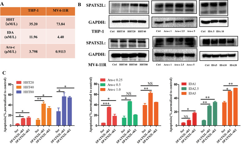
Background: Spermatogenesis associated serine rich 2 like (SPATS2L) was highly expressed in homoharringtonine (HHT) resistant acute myeloid leukemia (AML) cell lines. However, its role is little known in AML. The present study aimed to investigate the function of SPATS2L in AML pathogenesis and elucidate the underlying molecular mechanisms.
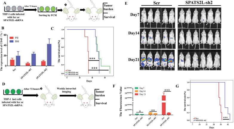
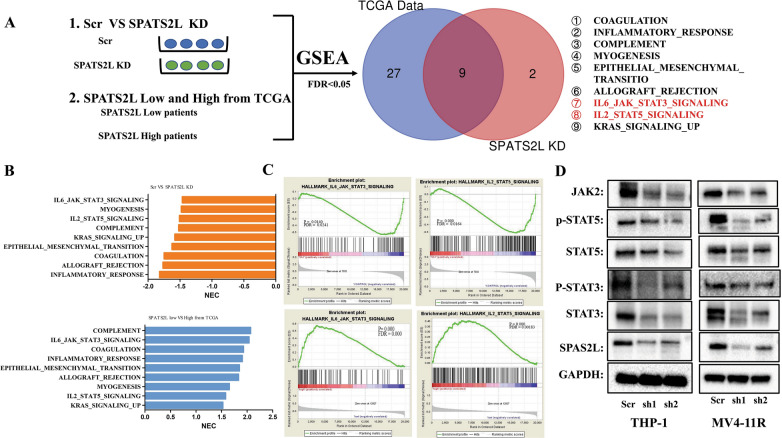
Methods: Overall survival (OS), event-free survival (EFS), relapse-free survival (RFS) were used to evaluate the prognostic impact of SPATS2L for AML from TCGA database and ourcohort. ShRNA was used to knockdown the expression of SPATS2L. Apoptosis was assessed by flow cytometry. The changes of proteins were assessed by Western blot(WB). A xenotransplantation mice model was used to evaluate in vivo growth and survival. RNA sequencing was performed to elucidate the molecular mechanisms underlying the role of SPATS2L in AML.
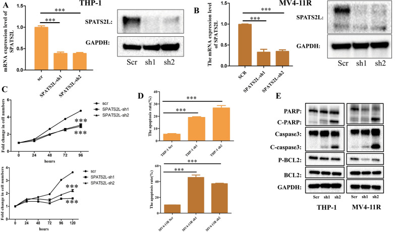
Results: SPATS2L expression increased with increasing resistance indexes(RI) in HHT-resistant cell lines we had constructed. Higher SPATS2L expression was observed in intermediate/high-risk patients than in favorable patients. Meanwhile, decreased SPATS2L expression was observed in AML patients achieving complete remission (CR). Multivariate analysis showed high SPATS2L expression was an independent poor predictor of OS, EFS, RFS in AML. SPATS2L knock down (KD) suppressed cell growth, induced apoptosis, and suppressed key proteins of JAK/STAT pathway, such as JAK2, STAT3, STAT5 in AML cells. Inhibiting SPATS2L expression markedly enhanced the pro-apoptotic effects of traditional chemotherapeutics (Ara-c, IDA, and HHT).
Conclusions: High expression of SPATS2L is a poor prognostic factor in AML, and targeting SPATS2L may be a promising therapeutic strategy for AML patients.
Keywords: AML; Apoptosis; Growth; JAK/STAT pathway; Prognostic; SPATS2L; Therapeutic target.
© 2023. The Author(s).
Conflict of interest statement
The authors declare that they have no competing interests.
Figures


Similar articles
-
Dihydropyrimidinase-like 2 can serve as a novel therapeutic target and prognostic biomarker in acute myeloid leukemia.Cancer Med. 2023 Apr;12(7):8319-8330. doi: 10.1002/cam4.5531. Epub 2023 Jan 9. Cancer Med. 2023. PMID: 36621846 Free PMC article.
-
Homoharringtonine affects the JAK2-STAT5 signal pathway through alteration of protein tyrosine kinase phosphorylation in acute myeloid leukemia cells.Eur J Haematol. 2008 Oct;81(4):259-66. doi: 10.1111/j.1600-0609.2008.01116.x. Epub 2008 Jul 8. Eur J Haematol. 2008. PMID: 18616510
-
Role of altered growth factor receptor-mediated JAK2 signaling in growth and maintenance of human acute myeloid leukemia stem cells.Blood. 2014 May 1;123(18):2826-37. doi: 10.1182/blood-2013-05-505735. Epub 2014 Mar 25. Blood. 2014. PMID: 24668492 Free PMC article.
-
JAKs to STATs: A tantalizing therapeutic target in acute myeloid leukemia.Blood Rev. 2020 Mar;40:100634. doi: 10.1016/j.blre.2019.100634. Epub 2019 Oct 25. Blood Rev. 2020. PMID: 31677846 Review.
-
The role of JAK/STAT signaling pathway in cerebral ischemia-reperfusion injury and the therapeutic effect of traditional Chinese medicine: A narrative review.Medicine (Baltimore). 2023 Nov 17;102(46):e35890. doi: 10.1097/MD.0000000000035890. Medicine (Baltimore). 2023. PMID: 37986307 Free PMC article. Review.
Cited by
-
RanBALL: An Ensemble Random Projection Model for Identifying Subtypes of B-Cell Acute Lymphoblastic Leukemia.bioRxiv [Preprint]. 2025 Jan 2:2024.09.24.614777. doi: 10.1101/2024.09.24.614777. bioRxiv. 2025. PMID: 39386448 Free PMC article. Preprint.
-
Homoharringtonine in the treatment of acute myeloid leukemia: A review.Medicine (Baltimore). 2024 Nov 1;103(44):e40380. doi: 10.1097/MD.0000000000040380. Medicine (Baltimore). 2024. PMID: 39496012 Free PMC article. Review.
-
A combination of Dihydroartemisinin and Venetoclax enhances antitumor effect in AML via C-MYC/BCL-XL/MCL-1 triple targeting.Discov Oncol. 2025 Apr 9;16(1):496. doi: 10.1007/s12672-025-02242-7. Discov Oncol. 2025. PMID: 40202582 Free PMC article.
-
Age-associated myeloid malignancies - the role of STAT3 and STAT5 in myelodysplastic syndrome and acute myeloid leukemia.FEBS Lett. 2024 Nov;598(22):2809-2828. doi: 10.1002/1873-3468.14985. Epub 2024 Jul 24. FEBS Lett. 2024. PMID: 39048534 Free PMC article. Review.
-
SPATS2L is a positive feedback regulator of the type I interferon signaling pathway and plays a vital role in lupus.Acta Biochim Biophys Sin (Shanghai). 2024 Aug 2;56(11):1659-1672. doi: 10.3724/abbs.2024132. Acta Biochim Biophys Sin (Shanghai). 2024. PMID: 39099414 Free PMC article.
References
-
- Yi M, Li A, Zhou L, Chu Q, Song Y, Wu K. The global burden and attributable risk factor analysis of acute myeloid leukemia in 195 countries and territories from 1990 to 2017: estimates based on the global burden of disease study 2017. J Hematol Oncol. 2020;13(1):72. doi: 10.1186/s13045-020-00908-z. - DOI - PMC - PubMed
Publication types
MeSH terms
Substances
LinkOut - more resources
Full Text Sources
Medical
Molecular Biology Databases
Miscellaneous
