. 2023 May 1:228:109455.
doi: 10.1016/j.neuropharm.2023.109455. Epub 2023 Feb 10.

Neuroimmune interactions with binge alcohol drinking in the cerebellum of IL-6 transgenic mice

Affiliations

Neuroimmune interactions with binge alcohol drinking in the cerebellum of IL-6 transgenic mice

Donna L Gruol et al. Neuropharmacology. .

Abstract

The neuroimmune system of the brain, which is comprised primarily of astrocytes and microglia, regulates a variety of homeostatic mechanisms that underlie normal brain function. Numerous conditions, including alcohol consumption, can disrupt this regulatory process by altering brain levels of neuroimmune factors. Alcohol and neuroimmune factors, such as proinflammatory cytokines IL-6 and TNF-alpha, act at similar targets in the brain, including excitatory and inhibitory synaptic transmission. Thus, alcohol-induced production of IL-6 and/or TNF-alpha could be important contributing factors to the effects of alcohol on the brain. Recent studies indicate that IL-6 plays a role in alcohol drinking and the effects of alcohol on the brain activity following the cessation of alcohol consumption (post-alcohol period), however information on these topics is limited. Here we used homozygous and heterozygous female and male transgenic mice with increased astrocyte expression of IL-6 to examined further the interactions between alcohol and IL-6 with respect to voluntary alcohol drinking, brain activity during the post-alcohol period, IL-6 signal transduction, and expression of synaptic proteins. Wildtype littermates (WT) served as controls. The transgenic mice model brain neuroimmune status with respect to IL-6 in subjects with a history of persistent alcohol use. Results showed a genotype dependent reduction in voluntary alcohol consumption in the Drinking in the Dark protocol and in frequency-dependent relationships between brain activity in EEG recordings during the post-alcohol period and alcohol consumption. IL-6, TNF-alpha, IL-6 signal transduction partners pSTAT3 and c/EBP beta, and synaptic proteins were shown to play a role in these genotypic effects.

Keywords: EEG; IL-6; STAT3; Signal transduction; Synapse; TNF-alpha; c/EBP beta; pSTAT3.


Conflict of interest statement

Declaration of competing interest The authors have no conflict of interest to declare.

Figures
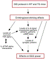
Figure 1.
Figure 1.
Schematic showing the areas of interest in the current studies. The studies focus on the neuroimmune factors IL-6 and TNF-alpha and IL-6 signal transduction partners (STAT3, c/EBP beta) that mediate the bioactivity of IL-6, and their role in alcohol drinking and brain activity during the post-alcohol period. Interactions between IL-6 and TNF-alpha and effects of neuroimmune/alcohol interactions on synaptic proteins that mediate brain activity also were examined. Arrows with solid lines indicate potential targets of alcohol/neuroimmune interactions that were studied. Arrow with dotted line indicates the expected target of the subject studied.
Figure 2.
Figure 2.
Water (ml/kg) and alcohol (EtOH)(g/kg) consumption in WT, +/−TG and +/+TG mice. A1,B1. Mean values (±SEM) for daily 2 hr (days 1-3) or 4 hr (day 4) water (A1) and alcohol (B1) consumption in mice subjected to the DID protocol. A2,B2. Mean values (±SEM) for total water (A2) and alcohol (B2) consumption during the 4 day test period. * = significantly different from WT of the same treatment group. Note difference in scale for figures A1 and B1.
Figure 3.
Figure 3.
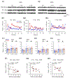
EEG power during the post-alcohol period in alcohol and water drinking mice. A1,2. Representative examples of EEG recordings in water (A1) and alcohol (A2) dinking WT, +/−TG and +/+TG mice during sleep stages that occurred during the EEG recording period. B1-3. Representative examples of hourly power for the delta band over the 20 hr EEG recording period in WT (B1), +/−TG (B2) and +/+TG (B3) mice. C1-4. Mean (±SEM) values for delta (C1), theta (C2), alpha (C3) and beta (C4) power during the 20 hr post-alcohol period in alcohol and water drinking WT, +/−TG and +/+TG mice. D1-3. Representative graphs of linear regression analysis for the relationship between power (alpha or theta) and day 4 alcohol consumption in WT (D1), +/−TG (D2) or +/+TG (D3) mice (see Supplemental Figure S1 for corresponding graphs of non-significant relationships for water consumption.) * = significantly different from WT of the same treatment groups (water and alcohol drinking data combined). @ = significant relationship.
Figure 4.
Figure 4.
Genotypic differences in cerebellar neuroimmune status of alcohol and water drinking WT, +/−TG and +/+TG mice. A,B. Mean (±SEM) values for IL-6 (A) and TNF-alpha (B) levels in cerebellum from WT, +/−TG and +/+TG mice. * = significantly different from WT (water and alcohol drinking data combined). C-E. Linear regression analysis of the relationship between cerebellar IL-6 and TNF-alpha levels in water or alcohol drinking WT (C1,C2), +/−TG (D1,D2) and +/+TG mice (E1,E2). F. Mean (±SEM) levels of STAT3 (F1), pSTAT3 (F2) and c/EBP beta (F3) in cerebellum from water and alcohol drinking WT, +/−TG and +/+TG mice. Representative Western blots are shown above the bars. Complete Western blots are shown in Supplemental Figure 4A. In this and other representative Western blots, the 42kD band is for beta actin. *= significantly different from WT of the same treatment group. & = significantly different from water drinking of the same genotype. @ = significant relationship.
Figure 5.
Figure 5.
Cerebellar GABAAR subunit expression in water and alcohol drinking mice. A. Mean (±SEM) levels of GABAAR of alpha 1,5,6 and delta subunits in cerebellum from water and alcohol drinking WT, +/−TG and +/+TG mice. Representative Western blots are shown above the graphs. Complete Western blots are shown in Supplemental figure 4B. B. Linear regression analysis of cerebellar GABAAR subunit expression and cerebellar levels of c/EBP beta (B1) or pSTAT3 (B2). Graphs show combined data from all genotypes and treatments. Protein levels were determined by Western blot analysis. Units for Western blots in this and other figures are ‘relative intensity’. * = significantly different from WT of the same treatment group. & = significantly different from water drinking of the same genotype. @ = significant relationship.
Figure 6.
Figure 6.
Genotypic and treatment differences in cerebellar synaptic protein expression. A-E. Mean (±SEM) levels of GABAAR subunits (A), gephyrin (B), GAD65/67 (C), GluR1 (D) and PSD95 (E) in cerebellum from alcohol and water drinking WT, +/−TG and +/+TG mice. Representative Western blots are shown above the graphs. In this and other representative Western blots, the 42kD band is for beta actin. Complete Western blots are shown in Supplemental figure 1B. * = significantly different from WT of the same treatment group. & = significantly different from water drinking of the same genotype.

Similar articles

Cited by

References

    1. Abrahao KP, Salinas AG, Lovinger DM, 2017. Alcohol and the Brain: Neuronal Molecular Targets, Synapses, and Circuits. Neuron 96, 1223–1238. - PMC - PubMed
    1. Alfonso-Loeches S, Pascual-Lucas M, Blanco AM, Sanchez-Vera I, Guerri C, 2010. Pivotal role of TLR4 receptors in alcohol-induced neuroinflammation and brain damage. Journal of Neuroscience 30, 8285–8295. - PMC - PubMed
    1. Alvarez Cooper I, Beecher K, Chehrehasa F, Belmer A, Bartlett SE, 2020. Tumour Necrosis Factor in Neuroplasticity, Neurogenesis and Alcohol Use Disorder. Brain Plast 6, 47–66. - PMC - PubMed
    1. Andersen LM, Jerbi K, Dalal SS, 2020. Can EEG and MEG detect signals from the human cerebellum? Neuroimage 215, 116817. - PMC - PubMed
    1. Baude A, Molnar E, Latawiec D, McIlhinney RA, Somogyi P, 1994. Synaptic and nonsynaptic localization of the GluR1 subunit of the AMPA-type excitatory amino acid receptor in the rat cerebellum. Journal of Neuroscience 14, 2830–2843. - PMC - PubMed

Publication types