. 2023 Mar;299(3):103004.
doi: 10.1016/j.jbc.2023.103004. Epub 2023 Feb 10.

Structural basis of nirmatrelvir and ensitrelvir activity against naturally occurring polymorphisms of the SARS-CoV-2 main protease

Affiliations

Structural basis of nirmatrelvir and ensitrelvir activity against naturally occurring polymorphisms of the SARS-CoV-2 main protease

Gabriela Dias Noske et al. J Biol Chem. 2023 Mar.

Abstract

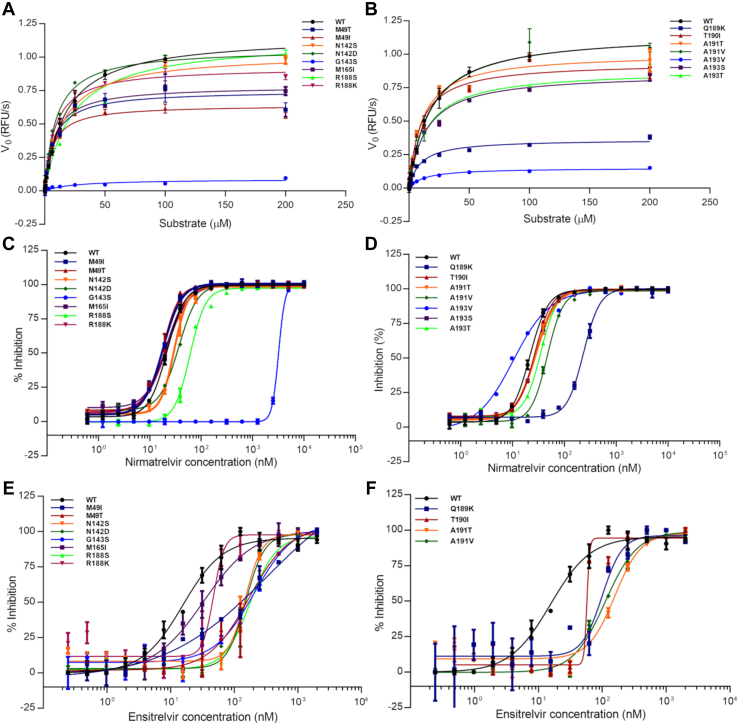
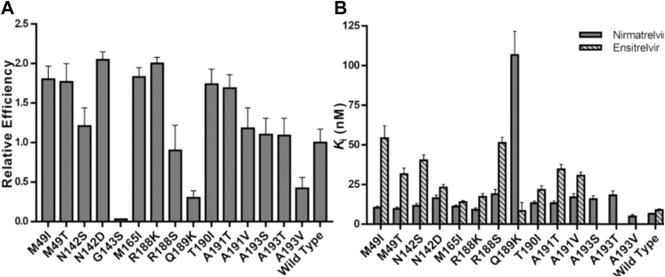
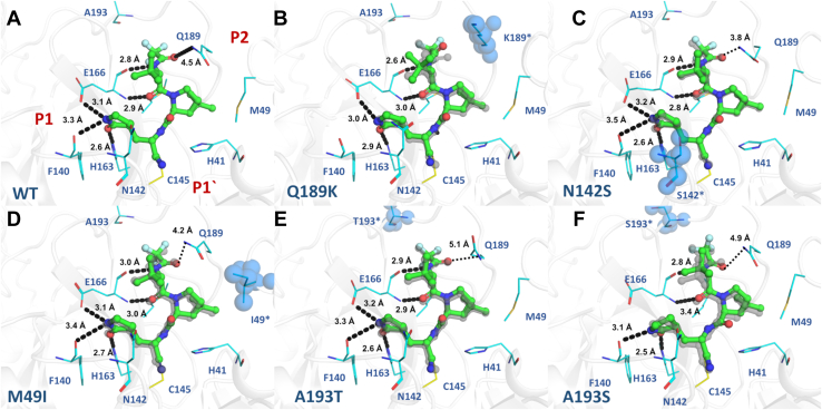
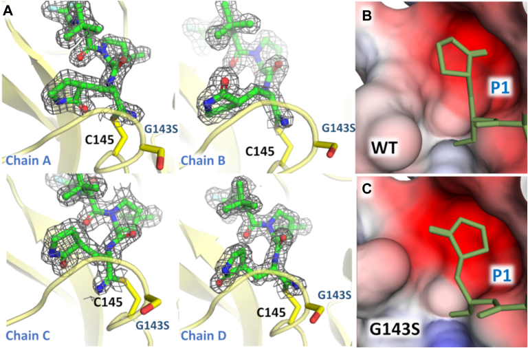
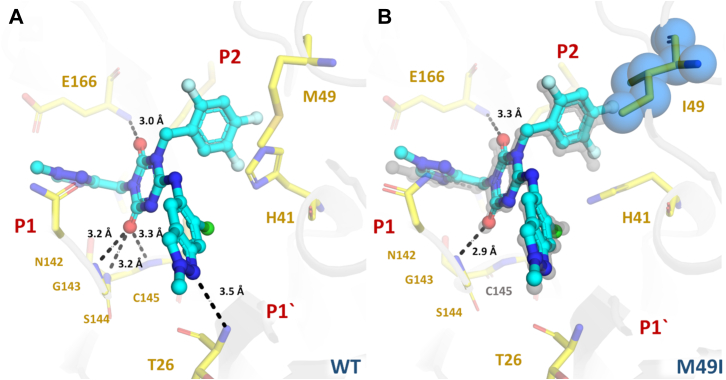
SARS-CoV-2 is the causative agent of COVID-19. The main viral protease (Mpro) is an attractive target for antivirals. The clinically approved drug nirmatrelvir and the clinical candidate ensitrelvir have so far showed great potential for treatment of viral infection. However, the broad use of antivirals is often associated with resistance generation. Herein, we enzymatically characterized 14 naturally occurring Mpro polymorphisms that are close to the binding site of these antivirals. Nirmatrelvir retained its potency against most polymorphisms tested, while mutants G143S and Q189K were associated with diminished inhibition constants. For ensitrelvir, diminished inhibition constants were observed for polymorphisms M49I, G143S, and R188S, but not for Q189K, suggesting a distinct resistance profile between inhibitors. In addition, the crystal structures of selected polymorphisms revealed interactions that were critical for loss of potency. In conclusion, our data will assist the monitoring of potential resistant strains, support the design of combined therapy, as well as assist the development of the next generation of Mpro inhibitors.

Keywords: COVID; SARS-CoV-2; drug; ensitrelvir; main protease; mpro; mutation; nirmatrelvir; paxlovid; resistance; structure; x-ray; xocova.


Conflict of interest statement

Conflict of interest The authors declare that they have no conflicts of interest with the contents of this article.

Figures

Figure 1
Figure 1
SARS-CoV-2 Mproamino acids polymorphisms identified in the genomic database.A, Mpro model showing variant spots identified in the genomic database, using a threshold of n ≥ 10. The Cα of identified variants spots are shown as cyan spheres, while red spheres are those variants within a radius of 8 Å ligands. Orange spheres show spots for variants of concern. Active site of Mpro is colored with a green blob. B, 2D plot of nirmatrelvir showing the distance and closest contacts with selected polymorphisms. C, 2D plot of ensitrelvir showing the distance and closest contacts with selected polymorphisms. D, panel containing polymorphism name, and substitution, and number of individuals identified (n) in a pool of 7 million SARS-CoV-2 genomes cataloged at CoV-GLUE by December 2021.
Figure 2
Figure 2
SARS-CoV-2 Mpropolymorphisms kinetics and inhibitions plots.A, Michaelis–Menten plots of WT and mutants M49T, M49I, N142S, N142D, G143S, M165I, R188S, and R188K. B, Michaelis–Menten plots of WT and mutants Q189K, T190I, A191T, A191V, A193V, A193S, and A193T. C, IC50 plots determination of nirmatrelvir against WT and mutants M49T, M49I, N142S, N142D, G143S, M165I, R188S, and R188K. D, IC50 plots determination of nirmatrelvir against WT and mutants Q189K, T190I, A191T, A191V, A193V, A193S, and A193T. E, IC50 plots determination of ensitrelvir against WT and mutants M49T, M49I, N142S, N142D, G143S, M165I, R188S, and R188K. F, IC50 plots determination of ensitrelvir against WT and mutants Q189K, T190I, A191T, and A191V. Data in graphs are means ± SD.
Figure 3
Figure 3
In vitro characterization of SARS-CoV-2 Mpromutants.A, relative catalytic efficiencies of Mpro mutants against FRET substrate. Data in graphs are means ± SD; n = 3. B, Ki determination of nirmatrelvir and ensitrelvir against Mpro mutants. Values were calculated using the results obtained in the FRET-based activity assay. For a better visualization, Ki values of mutant G143S are not represented in this graph. Data in graphs are means ± SD. FRET, fluorescence resonance energy transfer.
Figure 4
Figure 4
Crystal structures of Mpromutants in complex with nirmatrelvir. Mpro is displayed as cartoon and colored in gray; mutant residues are shown as spheres and colored in blue. Nirmatrelvir is shown as ball and stick and colored in green. Selected residues are shown as lines and colored in cyan. Substrate-binding subsites are labeled in red. A, Mpro WT (PDBid 8DZ2). B, mutant Q189K (PDBid 8DZ6). C, mutant N142S (PDBid 8E26). D, mutant M49I (PDBid 8E25). E, mutant A193T (PDBid 8DZA). F, mutant A193S (PDBid 8E1Y). WT crystal structure is aligned with each Mpro mutant, displayed as gray transparent sticks. Polar contacts are showed as black dashes.
Figure 5
Figure 5
Crystal structure of G143S Mpromutant in complex with nirmatrelvir.A, chains A–D of G143S Mpro in complex in nirmatrelvir highlighting the different ligand conformations. Mpro is shown as yellow cartoon. Residues G143 and C145 are shown as sticks and colored in yellow. For nirmatrelvir, the 2mFo-DFc electron density contoured at 1.0σ is represented in gray. B, surface charge representation of S1 subpocket for WT Mpro, showing the conformation of the pyrrolidine group. C, electrostatic charge calculated with APBS (43), projected on surface representation of S1 subpocket for G143S Mpro showing the conformation of the pyrrolidine group. For (B) and (C) nirmatrelvir is shown as lines and colored in yellow.
Figure 6
Figure 6
Crystal structures of Mproin complex with ensitrelvir. Mpro is displayed as cartoon and colored in gray; mutant residue is shown as spheres and colored in blue. Selected residues are displayed as yellow sticks. Ensitrelvir is shown as ball and stick and colored in cyan. Substrate-binding subsites are labeled in red. A, Mpro WT (PDBid 8DZ0). B, mutant M49I (PDBid 8DZ1) aligned with Mpro WT, displayed as gray transparent sticks. Polar contacts are showed as black dashes.

References

    1. Zhou P., Yang X.L., Wang X.G., Hu B., Zhang L., Zhang W., et al. A pneumonia outbreak associated with a new coronavirus of probable bat origin. Nature. 2020;579:270–273. - PMC - PubMed
    1. Wu F., Zhao S., Yu B., Chen Y.M., Wang W., Song Z.G., et al. A new coronavirus associated with human respiratory disease in China. Nature. 2020;579:265–269. - PMC - PubMed
    1. Jin Z., Du X., Xu Y., Deng Y., Liu M., Zhao Y., et al. Structure of Mpro from SARS-CoV-2 and discovery of its inhibitors. Nature. 2020;582:289–293. - PubMed
    1. Finkel Y., Mizrahi O., Nachshon A., Weingarten-Gabbay S., Morgenstern D., Yahalom-Ronen Y., et al. The coding capacity of SARS-CoV-2. Nature. 2020;589:125–130. - PubMed
    1. Lee J., Worrall L.J., Vuckovic M., Rosell F.I., Gentile F., Ton A.T., et al. Crystallographic structure of wild-type SARS-CoV-2 main protease acyl-enzyme intermediate with physiological C-terminal autoprocessing site. Nat. Commun. 2020;11:1–9. - PMC - PubMed

Publication types