[Preprint]. 2023 Jan 30:arXiv:2204.06159v2.

Systematic conformation-to-phenotype mapping via limited deep-sequencing of proteins

Affiliations

Systematic conformation-to-phenotype mapping via limited deep-sequencing of proteins

Eugene Serebryany et al. ArXiv. .

Update in

Abstract

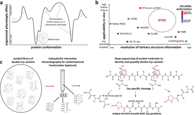
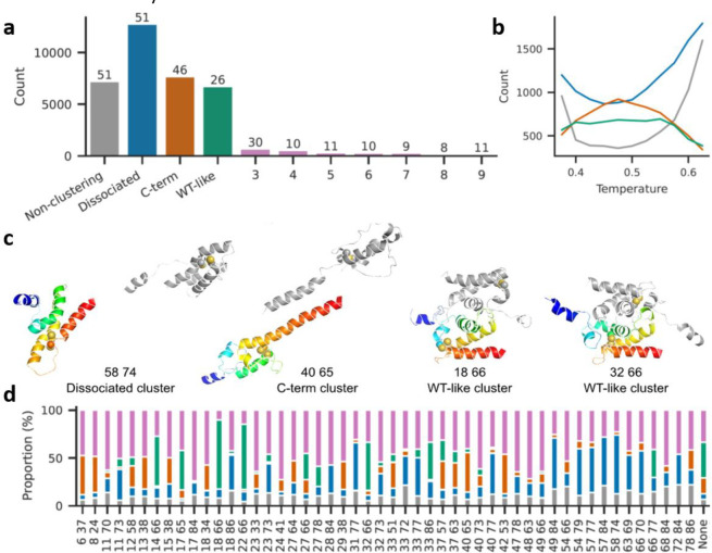
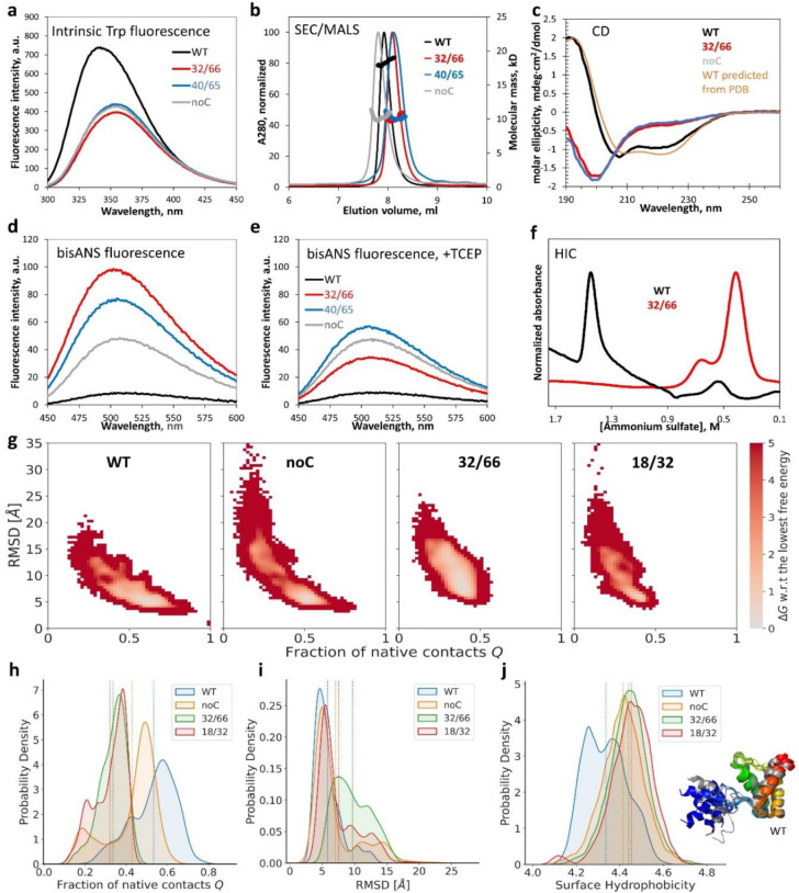
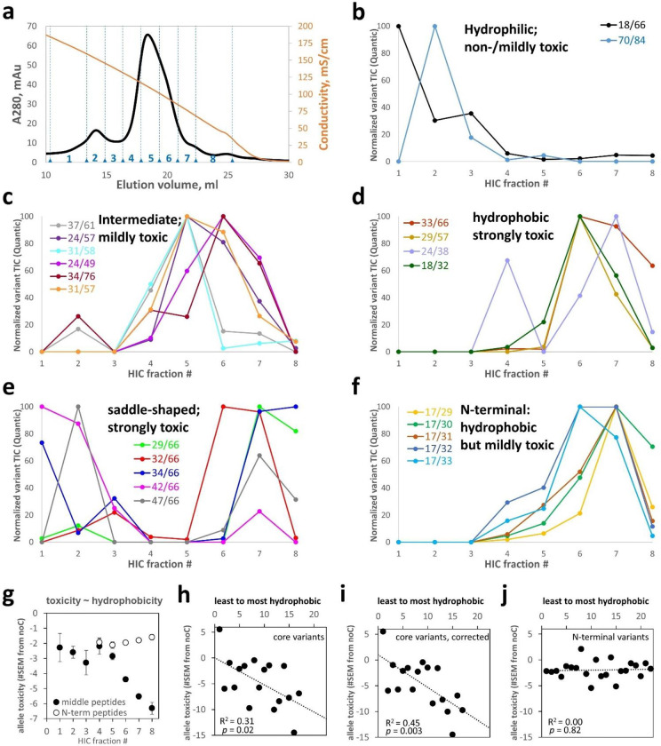
Non-native conformations drive protein misfolding diseases, complicate bioengineering efforts, and fuel molecular evolution. No current experimental technique is well-suited for elucidating them and their phenotypic effects. Especially intractable are the transient conformations populated by intrinsically disordered proteins. We describe an approach to systematically discover, stabilize, and purify native and non-native conformations, generated in vitro or in vivo, and directly link conformations to molecular, organismal, or evolutionary phenotypes. This approach involves high-throughput disulfide scanning (HTDS) of the entire protein. To reveal which disulfides trap which chromatographically resolvable conformers, we devised a deep-sequencing method for double-Cys variant libraries of proteins that precisely and simultaneously locates both Cys residues within each polypeptide. HTDS of the abundant E. coli periplasmic chaperone HdeA revealed distinct classes of disordered hydrophobic conformers with variable cytotoxicity depending on where the backbone was cross-linked. HTDS can bridge conformational and phenotypic landscapes for many proteins that function in disulfide-permissive environments.


Figures

Figure 1:
Figure 1:. Rationale and design for the HTDS method.
(a) In an IDP, distinct backbone conformations may produce distinct molecular and organismal phenotypes, such as cytotoxicity. (b) The niche for HTDS as a structural method: mapping both native and non-native conformers, both in vitro and in vivo, at moderate-to-high resolution. It is perhaps unique among all methods in its ability to connect specific conformational restraints to specific in vivo phenotypic effects. (c) The key enabling technology for HTDS is single-molecule deep sequencing of double-Cys protein libraries to locate both Cys in each polypeptide at once, thereby identifying and quantifying each variant across library fractions. We accomplish this via cyanylation-aminolysis: at pH 9, the amine of free Arg attacks the carbonyl immediately upstream of cyano-Cys – a good leaving group thanks to internal cyclization to iminothiazole (ITZ) – and is incorporated into the upstream fragment. The library may be fractionated by biophysical properties (e.g., hydrophobicity, as shown) or by chemical properties, such as redox status (i.e., disulfide-forming variants from non-disulfide-forming). Both approaches are demonstrated in this study.
Figure 2:
Figure 2:. HdeA variants exhibit varying levels of toxicity characterized by DnaK overexpression and cell lysis.
(a) Growth curves of HdeA variants in standard LB broth with varying [inducer], measured in parallel in the same 96-well plate, with all wells for each variant started from the same inoculum, showed variant- and [inducer]-dependent drops in OD600. WT HdeA and sGFP did not. (b) Non-reducing SDS-PAGE of end-point samples from 2500 μM rhamnose cultures showed clear expression of all constructs, albeit weaker than sGFP. Variants with more sequence-distal Cys pairs migrated lower on the gel, consistent with greater compaction of the unfolded state by longer-range disulfides (see Figure SI 6); note rightward skew of the gel. Juxtaposing identical volumes of centrifuged cell culture supernatant (“SUP”) and total culture (“TOT”) samples showed 86±11% (mean±S.D.) of HdeA in SUP across variants. All mutants showed many cytoplasmic protein bands in SUP, indicating cell lysis, and DnaK overexpression (confirmed by MS/MS). DnaK’s prominence in SUP indicated that it did not prevent lysis. (c) Non-reducing SDS-PAGE of the WT, 32/66, and noC constructs (TOT) as a function of [rhamnose]. (d) Quantitation of the HdeA bands in c, internally normalized to the abundant 37 kD band, showed a crossover between WT and 32/66 abundance. (e) Quantitation of the DnaK bands in panel c for 32/66 (orange line) and noC (light-blue line); observed [DnaK]/[HdeA] ratios converged to ~0.25.
Figure 3:
Figure 3:. Allele toxicity landscape of 1,453 double-Cys variants.
(a) A cartoon illustrating the experiment: HdeA variants were expressed in pooled culture; plasmids encoding lysogenic variants escaped into the culture medium as their host cells lysed, while those encoding less-toxic variants remained inside their host cells. During subsequent centrifugation, the former partitioned to the supernatant fraction, and the latter partitioned to the pellet. (b) An allele toxicity landscape for all 2-Cys variants that could be quantified by DNA deep sequencing in quadruplicate cultures grown with 1 mM rhamnose. Allele toxicity is defined as the difference of means of E between the given variant and noC, divided by the S.E.M. of the variant. Positive difference (orange) indicates less cell lysis than noC; negative difference (blue) indicates more lysis than noC. Very few variants were observed to be less toxic than noC. The WT gene (18/66, indicated by red arrow) ranked #3 out of 1,453 by this metric. (c) A pairwise contact map of Cα-Cα distances relative to WT’s, derived by averaging such distances in chain A and chain B of PDB ID 5WYO. (d) Cumulative average epistatic toxicity (expressed as in panel b) as a function of the pairwise native-state Cα-Cα distance (black) from WT for all double-Cys variants with Cys at least 12 peptide bonds apart showed lower cytotoxicity for smaller Cα-Cα distances. (e) Low-throughput measurements of expression levels of 38 randomly chosen double-Cys variants from the top and bottom 10% of the distribution of epistatic allele toxicities, defined as in panel (b).
Figure 4:
Figure 4:. Disulfide-bonding propensities of 166 double-Cys variants.
(a) Heatmap showing the abundance ratios (by total ion current) for variants found in NEM-treated (“PERI_NEM”) and untreated (“PERI”) periplasmic samples. Abundance of the most abundant variants found only without NEM is in gray. The green arrow indicates WT. Variants identified exclusively from N-terminal missed-cyanylation peptides are marked with green dots. The green box highlights variants containing Cys66, which were purposely enriched in the library. (b) Abundances in PERI and PERI_NEM (quantified by total ion current of the middle peptides, using Quantic) were strongly correlated, with a linear-regression trendline (black dashed line) close to the ratio measured for WT (red dashed line), so the vast majority of variants formed disulfides. Axes in thousands. (c) Same as b but for variants identified from N-terminal missed-cyanylation peptides only. (d) Violin plots of DNA-level allele abundance distributions (log-scale) of HdeA double-Cys variants detected vs. not detected at the protein level in the dataset in panel (a) illustrate that protein variants whose alleles were enriched at the DNA level in the library were much more likely to be detected in MS/MS. Only the theoretically detectable variants (Cys residues 11–62 peptide bonds apart) are considered.
Figure 5:
Figure 5:. Many non-native disulfides cause dissociation of HdeA dimers and melting of monomers in atomistic simulations.
(a) Combined sizes of top 10 clusters from all variant simulations; the number above each bar indicates how many variants had structures in that cluster. Total structures: 61600. Total clusters: 1032. Total structures belonging to none of the three main clusters: 29455. (b) Variation in cluster sizes with simulation temperature (colors as in a). (c) Representative structures from the three main clusters (numbers indicate Cys positions). The A subunit is in rainbow colors while the B subunit is gray. Cysteine residues are shown as spheres. Simulations included a 13-residue Gly-Ser linker between the subunits, here omitted for clarity. (d) Proportional cluster sizes for each simulated variant (colors as in a). “None” signifies the noC variant.
Figure 6:
Figure 6:. Biophysical characterization indicates HdeA mutants are monomeric, highly disordered, and hydrophobic.
(a) Intrinsic tryptophan fluorescence was quenched and red-shifted in 32/66, noC, and 40/65 compared to WT. (b) SEC/MALS revealed that, despite similar elution positions, only WT was dimeric; all mutants were monomers. (c) CD of WT matched predictions from PDB ID 5WYO by the PDB2CD tool (https://pdb2cd.cryst.bbk.ac.uk), but 32/66 and noC clearly did not. (d) The hydrophobicity probe bisANS bound much more strongly to the mutants (same samples as in a), especially 32/66. (e) Upon reduction of the disulfides, 32/66 and 40/65 bisANS fluorescence resembled noC; WT was resistant to reduction. (f) Folded (WT) and disordered (32/66) were easily separated by HIC. (g) Calculated free energy landscapes of monomeric WT, noC, and two double-Cys variants from MCPU simulations; the monomeric WT starting structure is defined as Q = 1. (h) The distribution of Q-values (fraction of native contacts) for mutant and WT HdeA. (i) The corresponding distribution of RMSD values relative to the starting WT structure. (j) The distribution of surface hydrophobicity values for WT dimer and mutant monomers; although it appears bimodal for WT, representative structures of the WT subunits (rainbow- vs. gray-colored ribbon diagrams) did not appreciably differ outside the N-terminal disordered region (blue/gray), which was often helical in our simulations. Dashed lines in panels h-j are distribution averages.
Figure 7:
Figure 7:. HIC of a pooled HdeA variant library reveals a correlation between hydrophobicity and toxicity.
(a) HIC elution trace of the total HdeA variant library on an ammonium sulfate gradient reported by conductivity (orange curve). Eight fractions were collected, numbered as indicated. Elution profiles of all 17 “core” variants are shown, grouped by similarity of elution profiles and centroids of elution: (b) below 4.7, (c) 4.7–6.0, (d) 6.0–6.8. (e) Some toxic core variants had unusual saddle-shaped elution profiles. (f) Most non-core variants were identified from N-terminal peptides, many with the missed cyanylation at position 17; those variants had high hydrophobicity despite modest toxicity. (g) Hydrophobicity and toxicity (PSM-averaged E value) were strongly correlated for variants identified from middle peptides but not from N-terminal peptides. (h) Differences between core and N-terminal variants accounted for this divergence, especially after correcting for centroid shifts due to saddle-shaped elution profiles (by ignoring data from the first 3 HIC fractions) (i). (j) By contrast, non-core variants – identified from either set of peptides – showed no hydrophobicity-toxicity correlation.

Similar articles

References

    1. Uversky V.N. (2019). Intrinsically Disordered Proteins and Their “Mysterious” (Meta)Physics. Front. Physics 7, 18, 10. 10.3389/fphy.2019.00010. - DOI
    1. Porter L.L., and Looger L.L. (2018). Extant fold-switching proteins are widespread. Proceedings of the National Academy of Sciences of the United States of America 115, 5968–5973. 10.1073/pnas.1800168115. - DOI - PMC - PubMed
    1. Brockwell D.J., and Radford S.E. (2007). Intermediates: ubiquitous species on folding energy landscapes? Current Opinion in Structural Biology 17, 30–37. 10.1016/j.sbi.2007.01.003. - DOI - PMC - PubMed
    1. Borgia A., Kemplen K.R., Borgia M.B., Soranno A., Shammas S., Wunderlich B., Nettels D., Best R.B., Clarke J., and Schuler B. (2015). Transient misfolding dominates multidomain protein folding. Nature Communications 6, 8861. 10.1038/ncomms9861. - DOI - PMC - PubMed
    1. Ascenzi P., and Gianni S. (2013). Functional Role of Transient Conformations: Rediscovering “Chronosteric Effects” Thirty Years Later. Iubmb Life 65, 836–844. 10.1002/iub.1208. - DOI - PubMed

Publication types

LinkOut - more resources