. 2023 Jan 26:14:986175.
doi: 10.3389/fimmu.2023.986175. eCollection 2023.

Secreted glucose regulated protein78 ameliorates DSS-induced mouse colitis

Affiliations

Secreted glucose regulated protein78 ameliorates DSS-induced mouse colitis

Liang Zhao et al. Front Immunol. .

Abstract

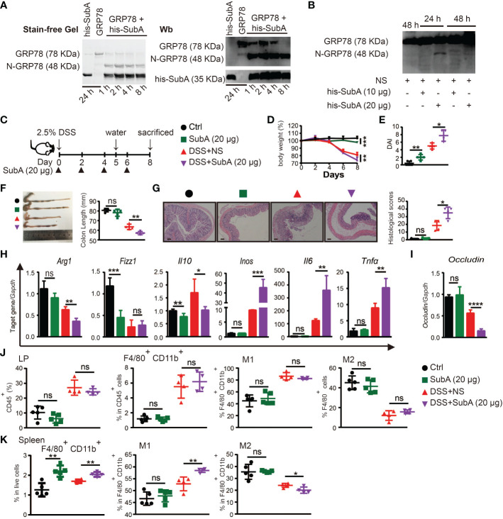
The secreted form of 78-kDa glucose-regulated protein (sGRP78) has been widely reported for its property in aiding resolution of inflammatory. However, little is known on its potential in the treatment of colitis. To investigate the expression pattern and functional outcome of GRP78 in ulcerative colitis, its expression was measured in human and murine colitis samples. It was found that GRP78 was spontaneously secreted to a high level in gut, which is a physiological site of immune tolerance. During the active phase of DSS-induced colitis, the sGRP78 level was significantly reduced but rebounded quickly during resolving phase, making it a potential candidate for the treatment of colitis. In the following experiments, the administration of sGRP78 was proved to decrease susceptibility to experimental colitis, as indicated by an overall improvement of intestinal symptoms, restoration of TJ integrity, decreased infiltration of immune cells and impaired production of inflammatory cytokines. And specific cleavage of endogenous sGRP78 could aggravate DSS colitis. Adoptive transfer of sGRP78-conditioned BMDMs reduced inflammation in the gut. We linked sGRP78 treatment with altered macrophage biology and skewed macrophage polarization by inhibiting the TLR4-dependent MAP-kinases and NF-κB pathways. Based on these studies, as a naturally occurring immunomodulatory molecule, sGRP78 might be an attractive novel therapeutic agent for acute intestinal inflammation.

Keywords: DSS; immunomodulatory; macrophage; sGRP78; ulcerative colitis.


Conflict of interest statement

The authors declare that the research was conducted in the absence of any commercial or financial relationships that could be construed as a potential conflict of interest.

Figures

Figure 1
Figure 1
GRP78 is decreased in the intestinal mucosa of UC patients. (A) Representative immunofluorescent staining of GRP78 in human colonic tissues (left). Scare bar: 50 μm. The quantitative results about the percentage of GRP78 immunoreactive cells per field of view was shown in right. sGRP78 (B) and TNF-α (C) levels in the supernatants of mucosa punches collected from UC (n=8) and non-colitic control (n=6) patients. The levels were normalized to colonic surface area. Results are expressed as mean ± standard error of mean (SEM). ns, not significant. *p < 0.05, **p < 0.01.
Figure 2
Figure 2
sGRP78 expression in experimental models of colitis. (A) Scheme of colitis induction. (B) Body weight was measured every two days and expressed as the percentage from day 0. At indicated time points, the colonic tissues were removed from sacrificed mice for following measurements: (C) Colon length. (D) Histological scores. Levels of TNF-α (E) and sGRP78 (F) in colonic punches supernatants. (G) Correlation analysis between sGRP78 and TNF-α (n=39). Pearson correlation coefficients (r) and p-values were calculated and shown. Relationship curve of sGRP78 with weight loss (H), colon shortening (I), and histological changes (J) in colitis. (K) Representative photomicrographs of colon sections (left) stained with HE (upper panel) or GRP78 (lower panel). (Day 0: black circle; Day2: red square; Day4: blue regular triangle; Day6: Purple inverted triangle; Day8: orange rhombus; Day10: black pentagram; Day12: brown error). Scale bar: 100 μm (upper panel) and 50 μm (lower panel). Numbers of GRP78-positive cells were calculated and shown in the right. Data represent mean ± SEM (n=5–6). ns, not significant. *p < 0.05, **p < 0.01, ***p < 0.001, ****p < 0.0001.
Figure 3
Figure 3
sGRP78 mitigates DSS-induced colitis and effects of sGRP78 on cytokine profile. (A) Timetable scheme showing DSS and sGRP78 administration in mice. Effect of sGRP78 on (B) weight loss, (C) DAI score, (D) survival rate (n=10) in DSS exposed mice. At the end of observation (day 8 of experiments), mice were sacrificed and the colonic tissues were removed for following measurements: (E) Representative gross picture of colons (left) and quantification of colon length (right). (F) Representative HE-staining (left) and respective colitis scores of indicated groups (right). Scale bar: 100 μm. (G) Representative immunofluorescence images of occludin. (H) Occludin and (I) Cldn4 levels measured by WB and RT-qPCR. (J) FITC-dextran assay. (K) Frequencies of CD45+ cells in single-cell suspension of colon tissues. Results are expressed as the mean ± SEM (n=5 except figure D). *p<0.05, **p<0.01. Black circle stands for vehicle control, red square for DSS+NS group and blue triangle for DSS+sGRP78 group. (L) Levels of cytokines in colonic tissues culture supernatant. (M) mRNA expression of Tnfα, Il6 and Il10 in colonic tissues. (N) Plasma concentrations of cytokines. IL-6 and TNF-α were measured by CBA and IL-10 by ELISA. (O) Western blot analysis of the expression of TLR4, iNOS, COX2, (P) MAPKs and p65 in colonic tissues. Data represent mean ± SEM (n=5). *p < 0.05, **p < 0.01, ***p < 0.001, ****p < 0.0001.
Figure 4
Figure 4
sGRP78 induced macrophage M2 polarization in colitis. Representative immunofluorescent images of GRP78 (green) and CD68 (red) stained human (A) and mouse (B) colonic mucosa. Scale bar: 50 μm. (C) Frequencies of F4/80+ cells in lamina propria (LP) and their CD80+ or CD206+ expressions. Upper: gating strategy followed to distinguish the LP macrophages. Middle: representative FCM dot plots. Lower: FCM plots based quantitative analysis for F4/80+CD11b+ macrophages. (D) Frequencies of indicated cell populations in spleen. (E) Frequencies of indicated cell populations in MLN. (F) mRNA expression of Arg1, Fizz1, Ym1, Mgl1, and Inos in mice colonic tissues (Ctrl group: black circle; DSS+NS group: red square; DSS+GRP78 group: blue regular triangle). Data represent mean ± SEM (n=5). ns, not significant. *p < 0.05, **p < 0.01, ****p < 0.0001.
Figure 5
Figure 5
sGRP78 reverses LPS-induced macrophage M1 polarization in vitro. (A) Binding of recombinant mouse sGRP78 with F4/80+ BMDMs. Scale bar: 20 μm. (B) Representative photographs of BMDMs conditioned by sGRP78 (10 μg/ml) or LPS (100 ng/ml) for 24 h (magnification 100×). Scale bar: 100 μm. (C) Levels of Arg1, Tnfα, Il6 and Inos in peritoneal macrophages. Western blot analysis of the expression of MAPKs, p65 (D), and TLR4, iNOS, COX2 (E) of BMDMs. To further assess the effect of sGRP78 on LPS-induced macrophage, BMDMs were stimulated with LPS for 18 h and then with LPS or sGRP78 for another 12h (for mRNA detection F) or 24h (for cytokine release, G). (H) Levels of CD80 and CD206 on BMDMs. Error bars represent mean ± standard deviation from triplicate samples in one experiment. *p < 0.05, **p < 0.01, ***p < 0.001, ****p < 0.0001.
Figure 6
Figure 6
Adoptive transfer of sGRP78-conditioned BMDMs ameliorated DSS-induced colitis. (A) Timetable scheme showing DSS and BMDMs administration in mice. Effect of sGRP78-induced BMDMs on (B) weight loss, (C) DAI score (n=5) in DSS exposed mice. (D) Representative gross picture of colons (left) and quantification of colon length (right). (E) Representative HE-staining (left) and respective colitis scores of indicated groups (right). Scale bar: 100 μm. (F) mRNA expression of Arg1, Fizz1,Il10, Inos, Il6, Tnfα, Occludin in colonic tissues. (G) FITC-dextran concentrations in serum of mice. (Ctrl group: black circle; DSS+NS group: green square; DSS+BMDMs group: red regular triangle; DSS+BMDMs (GRP78) group: Purple inverted triangle). Data represent mean ± SEM (n=4-5). ns, not significant. *p < 0.05, **p < 0.01, ***p < 0.001.
Figure 7
Figure 7
Degradation of sGRP78 aggravates DSS-induced colitis. (A) In vitro cleavage of sGRP78 (20 μg/ml) by its selective serine proteinase subA. (B) Cleavage of endogenous sGRP78 by intraperitoneally injected subA. (C) Timetable scheme showing DSS and subA administration in mice and symbols of each group (Ctrl group: black circle; subA group: green square; DSS+NS group: red regular triangle; DSS+subA group: Purple inverted triangle). (D) Weight loss. (E) DAI and (F) representative gross picture of colons (left) and quantification of colon length (right). (G) Representative HE-stained colons and respective colitis scores of indicated groups. Scale bar: 100 μm. (H) mRNA expression of polarization-related genes in colonic tissues. (I) Colonic expression of Occludin measured by RT-qPCR. Frequencies of CD80+ (M1) or CD206+ (M2) cells in F4/80+ colonic LPLs (J) and spleen (K). Data represent mean ± SEM (n=4-5). ns, not significant. *p < 0.05, **p < 0.01, ***p < 0.001, ****p < 0.0001.

Similar articles

Cited by

References

    1. Bedoui Y, Guillot X, Selambarom J, Guiraud P, Giry C, Jaffar-Bandjee MC, et al. . Methotrexate an old drug with new tricks. Int J Mol Sci (2019) 20(20):5023. doi: 10.3390/ijms20205023 - DOI - PMC - PubMed
    1. Hodson R. Inflammatory bowel disease. Nature (2016) 540(7634):S97–S. doi: 10.1038/540S97a - DOI - PubMed
    1. Kaser A, Zeissig S, Blumberg RS. Inflammatory bowel disease. Annu Rev Immunol (2010) 28(1):573–621. doi: 10.1146/annurev-immunol-030409-101225 - DOI - PMC - PubMed
    1. Xavier RJ, Podolsky DK. Unravelling the pathogenesis of inflammatory bowel disease. Nature (2007) 448(7152):427–34. doi: 10.1038/nature06005 - DOI - PubMed
    1. Lavelle A, Sokol H. Gut microbiota-derived metabolites as key actors in inflammatory bowel disease. Nat Rev Gastroenterol Hepatol (2020) 17(4):223–37. doi: 10.1038/s41575-019-0258-z - DOI - PubMed

Publication types

Substances