. 2023 Jan 26:14:1076533.
doi: 10.3389/fphys.2023.1076533. eCollection 2023.

Virtual Fly Brain-An interactive atlas of the Drosophila nervous system

Affiliations

Virtual Fly Brain-An interactive atlas of the Drosophila nervous system

Robert Court et al. Front Physiol. .

Abstract

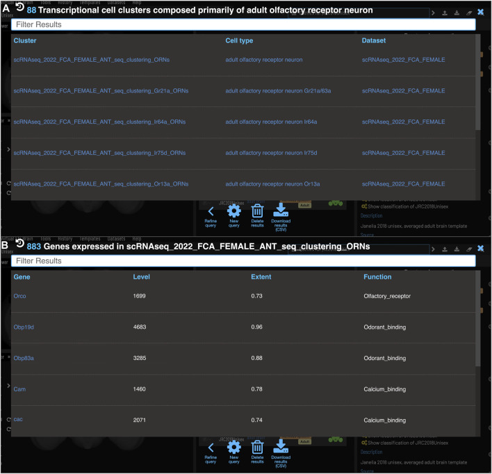
As a model organism, Drosophila is uniquely placed to contribute to our understanding of how brains control complex behavior. Not only does it have complex adaptive behaviors, but also a uniquely powerful genetic toolkit, increasingly complete dense connectomic maps of the central nervous system and a rapidly growing set of transcriptomic profiles of cell types. But this also poses a challenge: Given the massive amounts of available data, how are researchers to Find, Access, Integrate and Reuse (FAIR) relevant data in order to develop an integrated anatomical and molecular picture of circuits, inform hypothesis generation, and find reagents for experiments to test these hypotheses? The Virtual Fly Brain (virtualflybrain.org) web application & API provide a solution to this problem, using FAIR principles to integrate 3D images of neurons and brain regions, connectomics, transcriptomics and reagent expression data covering the whole CNS in both larva and adult. Users can search for neurons, neuroanatomy and reagents by name, location, or connectivity, via text search, clicking on 3D images, search-by-image, and queries by type (e.g., dopaminergic neuron) or properties (e.g., synaptic input in the antennal lobe). Returned results include cross-registered 3D images that can be explored in linked 2D and 3D browsers or downloaded under open licenses, and extensive descriptions of cell types and regions curated from the literature. These solutions are potentially extensible to cover similar atlasing and data integration challenges in vertebrates.

Keywords: FAIR; atlas; connectomics; drosophila; neurobiology; ontology; transcriptomics.


Conflict of interest statement

The authors declare that the research was conducted in the absence of any commercial or financial relationships that could be construed as a potential conflict of interest.

Figures

FIGURE 1
FIGURE 1
Templates and content. VFB has templates that integrate image data into common coordinate spaces and many more that serve as references for datasets in their native space The first four rows of the table provide details of the most up-to-date integrative templates on VFB: the JRC2018 unisex adult brain template (Bogovic et al., 2019) has the largest number of aligned images, which include over 26,500 EM images from CATMAID FAFB (Zheng et al., 2018) and the Janelia Hemibrain (Scheffer et al., 2020) combined, over 70,000 images showing expression patterns or fragments of expression patterns from sources including FlyCircuit (Chiang et al., 2010; Shih et al., 2015) and Janelia FlyLight (Meissner et al., 2022) and 46 painted neuropil domains (painted by Kazunori Shinomiya); The JRC2018 adult ventral nerve cord (VNC) template (Bogovic et al., 2019) has over 2,000 EM images from CATMAID FANC (Phelps et al., 2021), over 18,000 images of expression patterns from sources including FlyLight and 21 painted domains (Court et al., 2020) The Seymour L1 template has nearly 3,500 EM images from CATMAID L1 (Ohyama et al., 2015); The Wood2018 template has 255 painted domains (David Wood and Volker Hartenstein, unpublished). The rest of the table provides details of three of the available reference templated on VFB: the McKellar2020 adult head template has painted domains showing the adult pharyngeal musculature (McKellar et al., 2020); the hemibrain has the hemibrain connectome in its native space, along with a more detailed parcellation scheme (Scheffer et al., 2020); the Ito half-brain is the original reference template and parcellation scheme for the BrainName standard (Ito et al., 2014).
FIGURE 2
FIGURE 2
General layout and browsing. (A) The Slice Viewer allows users to view single slices of the Z-stack of the displayed elements. (B) The 3D Viewer shows entities in 3D space, allowing zoom and rotation. (C) The Template ROI Browser shows the neuropil regions of the current template (arranged hierarchically) and allows these to be added to the display. (D) The Layers tool acts as a color key for all the entities currently loaded and features a set of controls allowing content to be removed, hidden or recolored. (E) The term info shows details of a selected entity, in this case a cell type. Available images of this cell type are shown as thumbnails and can be added to the viewer by clicking the thumbnail. Split-GAL4 lines that target this cell type are also shown in the Term Info. Cell types also have a description based on published information. Term Info for a different entity can be shown by clicking on something in the Term Info or Layers panes, searching or using the left/right arrows above the Term Info pane. Arrowhead at top right indicates the search tool.
FIGURE 3
FIGURE 3
Search. Clicking the magnifying glass in the top right of the page will open the Search tool. Searching based on synonyms is supported and semantic tags on the right of each result provide extra information. Filters can be accessed by clicking on the lines on the right. (A) With no filters applied, results for “Or49a ORN” are a mixture of images (marked with *) and cell types from adult and larval stages. (B) To restrict results to larval neuron types, excluding images, filters can be applied to narrow down the results list, choosing a positive filter (green) for Larva and Neuron, and a negative filter (red) for Image.
FIGURE 4
FIGURE 4
Compound Queries. The Term Info pane (A) shows queries available in the TermInfo of “wedge projection neuron”. Clicking on the query for available images of “wedge projection neuron” bring up a results table (B) which can be further refined by clicking “Refine Query” underneath. The query interface (C,D) shows the original query and allows a second query to be run to find items that fit both sets of criteria, in this case images of neurons that also have some part in the lateral horn. Images in the subsequent results table (E) can be added to the viewer by clicking the checkboxes on the right.
FIGURE 5
FIGURE 5
Typing neurons using NBLAST. (A) TermIinfo for a neuron from FlyCircuit (Chiang et al., 2010) with no curated type other than “neuron”. (B) NBLAST query results for neurons similar morphology to the untyped query neuron. The top five results are all typed as “adult wedge projection neuron 2”. (C) image comparing the morphology of the query FlyCircuit neuron [“Cha-F-600036 (VFB_00007511)” in green] and the ‘adult wedge projection neuron 2' “WEDPN2B_R (FlyEM-HB:916828438) [VFB_jrchk7yi]" WEDPN2B_R in magenta).
FIGURE 6
FIGURE 6
Identifying Split-GAL4 combinations that potentially target a query neuron. (A) TermInfo for a neuron, “WEDPN2B_R”, type “adult wedge projection neuron 2” (WEDPN2), from the hemibrain dataset. This neuron will be used for an NBLAST search. (B) NBLAST query results showing Split-GAL4 driver line results and NBLAST scores. The third result (with the checked tickbox) was selected for further investigation (panels C and D). (C) Image of the query neuron skeleton (yellow) and Split-GAL4 expression pattern point cloud (blue) overlap. (D) A search for neuron types that this Split-GAL4 combination is known to target, curated from the literature, finds the type of the neuron used for the NBLAST search (WEDPN2), supporting the NBLAST query result in this case.
FIGURE 7
FIGURE 7
Identifying potential GAL4 drivers using color depth MIP scores (A) query results for the neuron “WEDPN2B_R”, showing hits to multiple MCFO images of driver line results and color depth MIP scores. The second (sparse MCFO expression) and fourth (dense MCFO expression) results (checked tickboxes) were selected for further investigation (panels (B) and (C), respectively). (B) Image of the query neuron (green) and expression pattern point cloud from a sparse line (magenta) overlap. (C) The same query neuron (green) also overlaps with the expression pattern point cloud of a dense line (magenta).
FIGURE 8
FIGURE 8
Circuit browser. A circuit diagram of paths between “WEDPN2B_R” and “VP2_adPN_R”. Rectangles represent neurons with the symbols of classes at the top, names of individual neurons at the bottom and colors corresponding to gross classifications in the middle. The legend for these gross classifications can be seen in the top-right [note the WDPN2 is classed as both cholinergic and glutamatergic based on antibody staining evidence (Dolan et al., 2019)]. Pathways are ordered from “strongest” at the bottom to “weakest” at the top. Arrows show the direction of synaptic connectivity and numbers outside of brackets show the number of synapses annotated for each connection. Numbers inside brackets show the number of synapses in the opposite direction.
FIGURE 9
FIGURE 9
Single Cell RNAseq (not yet live) (A) Each transcriptional cluster is linked to a cell type in the Drosophila Anatomy Ontology (curation done by Single Cell Expression Atlas and FlyBase) facilitating searches based on cell type (typically more general types than we have for connectomics data). (B) Each gene expressed in more than half of the cells in a cluster will be viewable in VFB with its expression level and extent (proportion of cells in cluster that transcript was detected in) and semantic tags representing the gene’s function (based on GO and Gene Group annotations from FlyBase).

References

    1. Bates A. S., Janssens J., Jefferis G. S., Aerts S. (2019). Neuronal cell types in the fly: Single-cell anatomy meets single-cell genomics. Curr. Opin. Neurobiol. 56, 125–134. 10.1016/j.conb.2018.12.012 - DOI - PubMed
    1. Bates A. S., Manton J. D., Jagannathan S. R., Costa M., Schlegel P., Rohlfing T., et al. (2020b). The natverse, a versatile toolbox for combining and analysing neuroanatomical data. eLife 9, e53350. 10.7554/eLife.53350 - DOI - PMC - PubMed
    1. Bates A. S., Schlegel P., Roberts R. J. V., Drummond N., Tamimi I. F. M., Turnbull R., et al. (2020a). Complete connectomic reconstruction of olfactory projection neurons in the fly brain. Curr. Biol. CB 30 (16), 3183–3199. 10.1016/j.cub.2020.06.042 - DOI - PMC - PubMed
    1. Bogovic J. A., Otsuna H., Heinrich L., Ito M., Jeter J., Meissner G., et al. (2019). An unbiased template of the Drosophila brain and ventral nerve cord. bioRxiv. 10.1101/376384 - DOI - PMC - PubMed
    1. Bunt S. M., Grumbling G. B., Field H. I., Marygold S. J., Brown N. H., Millburn G. H., et al. (2012). Directly e-mailing authors of newly published papers encourages community curation. Database J. Biol. databases curation 2012, bas024. 10.1093/database/bas024 - DOI - PMC - PubMed

LinkOut - more resources