. 2022 Nov 9;8(1):19-34.
doi: 10.1016/j.jacbts.2022.06.021. eCollection 2023 Jan.

1-Year Patency of Biorestorative Polymeric Coronary Artery Bypass Grafts in an Ovine Model

Affiliations

1-Year Patency of Biorestorative Polymeric Coronary Artery Bypass Grafts in an Ovine Model

Masafumi Ono et al. JACC Basic Transl Sci. .

Abstract

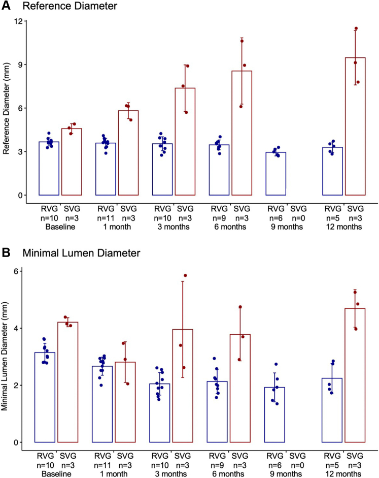
Many attempts have been made to inhibit or counteract saphenous vein graft (SVG) failure modes; however, only external support for SVGs has gained momentum in clinical utility. This study revealed the feasibility of implantation, and showed good patency out to 12 months of the novel biorestorative graft, in a challenging ovine coronary artery bypass graft model. This finding could trigger the first-in-man trial of using the novel material instead of SVG. We believe that, eventually, this novel biorestorative bypass graft can be one of the options for coronary artery bypass graft patients who have difficulty harvesting SVG.

Keywords: CABG, coronary artery bypass grafting; CPB, cardiopulmonary bypass; IH, intimal hyperplasia; LAD, left anterior descending artery; OCT, optical coherence tomography; QCA, quantitative coronary angiography; QFR, quantitative flow ratio; RVG, restorative vascular graft; SVG, saphenous vein graft; coronary artery bypass graft; coronary artery disease; coronary revascularization; ePTFE, expanded polytetrafluoroethylene; polymeric bypass graft; preclinical model; quantitative flow ratio; restorative vascular graft.


Conflict of interest statement

The work described in this paper was fully funded by Xeltis BV. Drs El-Kurdi, Reinöhl, and Cox are all employees of Xeltis. Dr Virmani has received personal fees from Xeltis, and Medtronic; as well as Institutional grants from Abbott Vascular, Boston Scientific, Medtronic, Xeltis and Becton Dickinson. Prof. Onuma has received institutional research grants related to his work as the Chairman of cardiovascular imaging core labs of several clinical trials and registries sponsored by industry, for which he receives no direct compensation. Prof. Serruys has received personal fees from Sino Medical Sciences Technology, Philips/Volcano and Xeltis. All other authors have reported that they have no relationships relevant to the contents of this paper to disclose.

Figures

None
Graphical abstract
Figure 1
Figure 1
Illustration of Implant Configuration and Implant Angiography A total of 16 sheep were used in this study: 13 were implanted with a restorative vascular graft and 3 with a saphenous vein graft. Only 1 graft was implanted per animal.
Figure 2
Figure 2
Image of RVG With Blow-Up to Show SEM Morphology A representative photograph of a restorative vascular graft (RVG) is presented in the left upper panel. A representative scanning electron photomicrograph of the outer surface of a device is shown in the lower left. The device exhibits a highly porous morphology characterized by bonded overlapping fibers ∼5 μm in diameter. RVG = restorative vascular graft; SEM = scanning electron microscopy.
Figure 3
Figure 3
Study Flowchart The study design and timeline are described in the schematic diagram. FU = follow-up; OCT = optical coherence tomography; RVG = restorative vascular graft; SVG = saphenous vein graft.
Figure 4
Figure 4
Serial Angiography Snapshot Images of All RVGs and SVGs in Animals That Survived to the 12-Month Follow-Up Representative snapshots from angiograms of all RVGs and SVGs in animals that survived to 12 months are presented. N/a = not applicable; other abbreviations as in Figure 3.
Figure 5
Figure 5
Serial Plots of Angiographic Parameters for All Animals Serial changes of reference diameter (A), minimal lumen diameter (B), flow speed (C), and quantitative flow ratio (QFR) (D) in both RVGs (blue) and SVGs (red). Bars are the SD of the mean. One occluded RVG (restorative vascular graft) (3-month follow-up) was not included in the graphs (19S0204).
Figure 5
Figure 5
Serial Plots of Angiographic Parameters for All Animals Serial changes of reference diameter (A), minimal lumen diameter (B), flow speed (C), and quantitative flow ratio (QFR) (D) in both RVGs (blue) and SVGs (red). Bars are the SD of the mean. One occluded RVG (restorative vascular graft) (3-month follow-up) was not included in the graphs (19S0204).
Figure 6
Figure 6
Series of All Measurements for Each Angiography-Derived Parameter Over Follow-Up Period Measurements are color-coded by group (RVG and SVG) with a smoothed average (locally estimated scatterplot smoothing) over time plotted for each group to illustrate group differences and changes over time. AS = area stenosis; DS = diameter stenosis; MLA = minimum lumen area; MLD = minimum lumen diameter; Ref. Diam. = reference diameter. Abbreviations as in Figures 3 and 5.
Figure 7
Figure 7
Representative Examples of Serial QFR in Cases of RVG and SVG This figure shows one representative case of an RVG and SVG, respectively, with serial QFR follow-ups out to 12 months. Abbreviations as in Figures 3 and 5.
Figure 8
Figure 8
Micro-CT Profile and Histologies at 12 Months Micro–computed tomography (CT) profile at 12-month explant of RVG (upper left) and SVG (lower left). (Right) Histology cross-sections near distal anastomosis of each graft (upper right, RVG; lower right, SVG). Abbreviations as in Figures 3 and 5.

References

    1. Taggart D.P., Ben Gal Y., Lees B., et al. A randomized trial of external stenting for saphenous vein grafts in coronary artery bypass grafting. Ann Thorac Surg. 2015;99:2039–2045. - PubMed
    1. Mack M.J., Squiers J.J., Lytle B.W., DiMaio J.M., Mohr F.W. Myocardial revascularization surgery: JACC historical breakthroughs in perspective. J Am Coll Cardiol. 2021;78:365–383. - PubMed
    1. Lytle B.W., Blackstone E.H., Sabik J.F., Houghtaling P., Loop F.D., Cosgrove D.M. The effect of bilateral internal thoracic artery grafting on survival during 20 postoperative years. Ann Thorac Surg. 2004;78:2005–2012. [discussion 2012-2014] - PubMed
    1. Ghista D.N., Kabinejadian F. Coronary artery bypass grafting hemodynamics and anastomosis design: a biomedical engineering review. Biomed Eng Online. 2013;12:129. - PMC - PubMed
    1. Ojha M., Cobbold R.S., Johnston K.W. Influence of angle on wall shear stress distribution for an end-to-side anastomosis. J Vasc Surg. 1994;19:1067–1073. - PubMed

LinkOut - more resources