. 2022 Aug 11:14:100340.
doi: 10.1016/j.lana.2022.100340. eCollection 2022 Oct.

E.L., a modern-day Phineas Gage: Revisiting frontal lobe injury

Affiliations

E.L., a modern-day Phineas Gage: Revisiting frontal lobe injury

Pedro H M de Freitas et al. Lancet Reg Health Am. .

Abstract

Background: How the prefrontal cortex (PFC) recovers its functionality following lesions remains a conundrum. Recent work has uncovered the importance of transient low-frequency oscillatory activity (LFO; < 4 Hz) for the recovery of an injured brain. We aimed to determine whether persistent cortical oscillatory dynamics contribute to brain capability to support 'normal life' following injury.

Methods: In this 9-year prospective longitudinal study (08/2012-2021), we collected data from the patient E.L., a modern-day Phineas Gage, who suffered from lesions, impacting 11% of his total brain mass, to his right PFC and supplementary motor area after his skull was transfixed by an iron rod. A systematic evaluation of clinical, electrophysiologic, brain imaging, neuropsychological and behavioural testing were used to clarify the clinical significance of relationship between LFO discharge and executive dysfunctions and compare E.L.´s disorders to that attributed to Gage (1848), a landmark in the history of neurology and neuroscience.

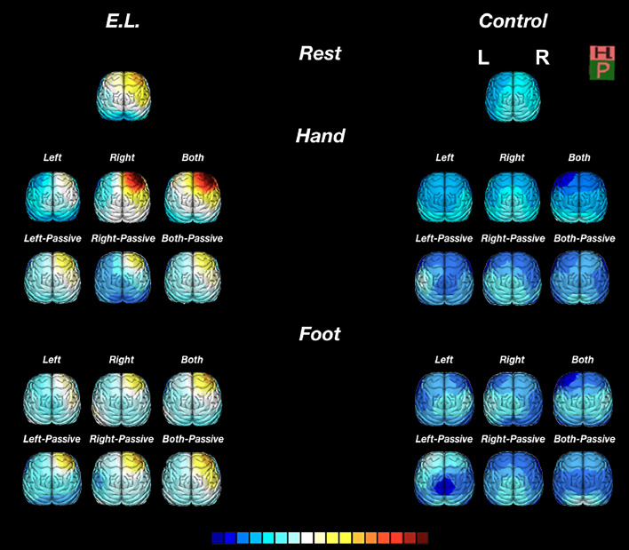
Findings: Selective recruitment of the non-injured left hemisphere during execution of unimanual right-hand movements resulted in the emergence of robust LFO, an EEG-detected marker for disconnection of brain areas, in the damaged right hemisphere. In contrast, recruitment of the damaged right hemisphere during contralateral hand movement, resulted in the co-activation of the left hemisphere and decreased right hemisphere LFO to levels of controls enabling performance, suggesting a target for neuromodulation. Similarly, transcranial magnetic stimulation (TMS), used to create a temporary virtual-lesion over E.L.'s healthy hemisphere, disrupted the modulation of contralateral LFO, disturbing behaviour and impairing executive function tasks. In contrast to Gage, reasoning, planning, working memory, social, sexual and family behaviours eluded clinical inspection by decreasing LFO in the delta frequency range during motor and executive functioning.

Interpretation: Our study suggests that modulation of LFO dynamics is an important mechanism by which PFC accommodates neurological injuries, supporting the reports of Gage´s recovery, and represents an attractive target for therapeutic interventions.

Funding: Fundação de Amparo Pesquisa Rio de Janeiro (FAPERJ), Universidade Federal do Rio de Janeiro (intramural), and Fiocruz/Ministery of Health (INOVA Fiocruz).

Keywords: C.C., Corpus callosum; Corpus callosum (C.C.); LFO, Low frequency oscillations (EEG); Low Frequency Oscillations; MRI, Magnetic Resonance Imaging; Magnetic Resonance Imaging (MRI); Neuropsychological tests; PFC, Prefrontal cortex; Phineas Gage; Prefrontal cortex (PFC); TBI, Traumatic brain injury; TMS, Transcranial magnetic stimulation; Transcranial Magnetic Stimulation; Traumatic brain injury (TBI).


Conflict of interest statement

All authors declare that they have no conflicts of interest.

Figures

Figure 1
Figure 1
Path of the iron bar through E.L.’s skull and its effects on white matter structure. (a) Lateral view of the transfixed skull with an iron bar on the CT scan reconstruction. (b) T1 weighted MRI reconstruction revealed the trajectory of the iron bar. Lateral (c) and transverse (d) sections (T2-weighted MRI images), 18 months after the accident, suggestive of a right frontal lobe disconnection; (e,g) FLAIR MRI spectroscopy (single-voxel) was applied to regions of interest (blue squares), indicated by blue arrows, on the anterior corpus callosum (CC). (f,h) An expected decrease in spectrum of resonances in N-acetyl-aspartate (NAA) (2ppm), marker of neuronal integrity, is illustrated. (i) Axial view of a tractography suggesting a difference in the volume of the association fiber tracts that connect temporo-parietal cortical regions with right frontal lobe (white arrow). (j) Fractional anisotropy (FA) map, a diffusion tensor imaging technique, revealed that the injured area was associated with greater anisotropy reduction in frontal right brain regions (white arrow). R (right hemisphere), L (left hemisphere).
Figure 2
Figure 2
E.L.: Figural fluency, emotional behaviour and EEG as a measure of right frontal lobe injury. This battery of tests was aimed at identifying dysfunction, such as attention and concentration, self-monitoring, personality, inhibition of behaviour and emotions, and with speaking or using expressive language. (a-d) Representative illustrations of memory for geometric designs, shapes, features and directional orientation. (a, top) E.L. was asked to drawn R$ 5 cents, 25 cents and 1 Real coins from memory (i.e., draw-to-command). Coin measurements were compared to the respective official coins (a, bottom). (e) The clock-drawing test did not confirm a diagnosis of cognitive deficits in E.L. Assessment focused on size of the clock, graphic difficulties, stimulus-bound response, conceptual deficit, spatial/planning deficit and perseveration. (f) context-dependence of emotions within text, illustrated by “Eu amo minha familia e meus pais” (“I love my family and my parents”). (g) Spontaneous EEG (four non-consecutive sec of the same recording session placed side by side). (i) Eye closure sensitivity: the awake EEG is characterized by a posterior dominant alpha rhythm (9-10Hz) and moderate voltage reactive to eye opening and closure. (ii) Awake, eyes-closed resting condition: there is frequent delta slowing seen in the right frontal region (Fp2-F4 and Fp2-F8). (iii) Hyperventilation (HV): HV showed a tendency to increase the focal slowing noted above. (iv) Photic Stimulation (PS): intermittent PS did not alter the background. Calibration bar: 100 µV.
Figure 3
Figure 3
Functional BOLD MRI images (fMRI) of E.L.’s brain activation during sequential finger-to-thumb tapping movements using one hand alone or two hands simultaneously. Axial (a-c), sagittal (d-f) and coronal sections (g-i) offer the possibility of directly investigating cortical brain activation and connectivity during task performance using the right hand alone (a,d,g), left hand alone (b,e,h) or during bimanual movements (c,f,i). Of note, use of either the left hand alone or simultaneously both hands showed a similar trend of activation in the bilateral frontal and parietal regions, known to be involved in working memory. By contrast, for the right-hand alone movements, activation of the motor control regions was lateralized to the left hemisphere. Scale: Z score ranging from -40.95 to 40.95.
Figure 4
Figure 4
Changes in slow wave amplitude of the quantitative EEG during active and passive (assisted) symmetric movement tasks. The topographic delta frequency band distribution in E.L. and CTRL (resting testing) (top row), during unimanual left/right and bimanual tasks (middle row) and during foot movements (bottom row) is illustrated. Significant differences from baseline for absolute amplitude (µV) in delta frequency were found only in E.L. right frontal region (Fp2) while performing active finger-to-thumb tapping movements – P < 0.05 baseline vs. left hand and P < 0.05 baseline vs. right hand. Changes in delta wave amplitude during active/passive hand or foot movements were neither documented for CTRL nor for E.L. during either active/passive foot and passive hand movements. Calibration (colour scale): 15-102 µV.
Figure 5
Figure 5
rTMS influences short-term memory and behaviour in E.L., but not in CTRL, by modulating the frequency of slow waves. (a) depicts the position of the conventional TMS figure-of-eight coil with respect to E.L.’s scalp, over the left motor cortex (M1). (b-d) T2- weighted imaging of E.L.’s brain from 2020, 8 years since injury. (b,d) TBI did not result in progressive loss of E.L.’s brain tissue volume. Mesial temporal sclerosis was not detected with regard to hippocampal size and shape, fissure visualization and signal intensity (isotense) to cortical gray matter. (e-h) rTMS-EEG responses to stimulation in E.L. and in CTRL. (e) Topographic plots of the TMS-evoked responses. The extent to which increase in frequency and amplitude of SWs are higher contralateral than ipsilateral to the side of stimulation is illustrated by a train of rTMS at a frequency of 1 Hz (top panel). After rTMS, EEG SW responses were potentiated up to 40 min post 1 Hz (f,h) and up to 60 min post-Theta-burst stimulation (TBS) (g,h), followed by a complete return to background activity (e,h – top panel). (i,j) Subjects were asked to copy the Rey–Osterrieth figure (bottom left – ‘ template’), and then to reproduce it from memory 3–30 min later, prior and after TMS-treatment (1 Hz) applied to the DLPFC. The two groups drawing scores significantly differ between the retrieval rounds at 3 min (* P < 0.001) and at 30 min (* P < 0.001). rTMS scale (e): 0–500 µV².

References

    1. Luria AR. Springer; New York, NY, USA: 1966. Higher Cortical Functions in Man.
    1. Harlow JM. Recovery from the passage of a iron bar through the head. Bost Med Surg J. 1868;2:327–346.
    1. Gazzaniga MS, Miller MB. The Neurology of Consciousness. Academic Press; San Diego, CA, USA: 2009. The left hemisphere does not miss the right hemisphere; pp. 261–270. - DOI
    1. Berlucchi G. Frontal callosal disconnection syndromes. CORTEX. 2011;48:36–45. - PubMed
    1. Gazzaniga MS. Cerebral specialization and interhemispheric communication does the corpus callosum enable the human condition? Brain. 2000;123:1293–1326. - PubMed