Review
. 2023 Jan 26:10:1037404.
doi: 10.3389/fvets.2023.1037404. eCollection 2023.

Periodic discharges in veterinary electroencephalography-A visual review

Affiliations
Review

Periodic discharges in veterinary electroencephalography-A visual review

Marguerite F Knipe et al. Front Vet Sci. .

Abstract

First described in human EEG over 60 years ago, there are very few examples of periodic discharges in the veterinary literature. They are associated with a wide variety of etiologies, both intracranial and systemic, making interpretation challenging. Whether these patterns are indicative of ictal, interictal, or postictal activity is a matter of debate and may vary depending on the clinical features in an individual patient. Periodic discharges have a repeated waveform occurring at nearly regular intervals, with varying morphology of individual discharges from simple sharp waves or slow waves to more complex events. Amplitudes, frequencies, and morphologies of the discharges can fluctuate, occasionally evolving, or resolving over time. This study presents a visual review of several veterinary cases with periodic discharges on EEG similar to those described in human EEG, and discusses the current known pathophysiology of these discharges.

Keywords: EEG; canine; encephalopathy; epilepsy; feline; seizures; status epilepticus.


Conflict of interest statement

The authors declare that the research was conducted in the absence of any commercial or financial relationships that could be construed as a potential conflict of interest.

Figures
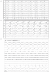
Figure 1
Figure 1
Three examples of generalized periodic discharges (GPDs). PDs are noted predominantly frontal and in both hemispheres in each case. (A) Ten year old mixed breed dog with several year history of seizures and recent cluster seizures. Polyphasic GPDs, synchronous and bilateral, with slightly higher amplitude in the left hemisphere (Bipolar montage; HF 70 Hz, sensitivity 5 uV/mm). (B) Four year old domestic longhair cat with otogenic bacterial encephalitis and seizures. Polyphasic GPDs (Bipolar montage; HF 70 Hz, sensitivity 7 uV/mm). (C) Thirteen year old Bichon frise with MUO and recent cluster seizures (Referential montage; HF 35 Hz, sensitivity 5 uV/mm). The amplitude of the discharge is slightly lower in F4 than F3 & Fz, although the timing of the PD is synchronous in the frontal leads. MUO, meningoencephalitis of unknown origin.
Figure 2
Figure 2
Referential (A–D) and bipolar (E) montages of five cases with lateralized periodic discharges (LPDs). (A) Left parietal LPDs in a 12 year old pug with cluster seizures, normal brain MRI (HF 35 Hz, sensitivity 7 uV/mm). (B) Frontal midline LPDs in a 14 year old Chihuahua mix with cluster seizures secondary to hypoglycemia and an insulin-secreting tumor (HF 30 Hz, sensitivity 10 uV/mm). (C) Right frontal LPDs in an 11 year old miniature Pinscher with recent SE and history of transfrontal craniotomy 6 months previously for right olfactory/frontal lobe meningioma (HF 35 Hz, sensitivity 15 uV/mm). (D) Left hemisphere LPDs in an 11 year old Labrador with recent cluster seizures and history of craniotomy 6 months previously for left occipital meningioma (HF 30 Hz, sensitivity 5 uV/mm). (E) Left hemisphere polyphasic LPDs (phase reversal at T3 and C3) in an 8 year old soft-coated Wheaton terrier with metastatic neoplasia (HF 35 Hz, sensitivity 5 uV/mm).
Figure 3
Figure 3
Four examples of periodic discharges, where the morphology of the PD (solid arrow) included a second or third waveform (arrowheads), with a consistent temporal relationship to the initial discharge, like a doublet or triplet. (A) Eleven year old Chihuahua with SE (Bipolar montage; HF 70 Hz, sensitivity 20 uV/mm). (B) Two year old bichon mix with progressive partial seizures and MUO (Bipolar montage; HF 70 Hz, sensitivity 10 uV/mm). (C) Three year old terrier mix with cluster seizures, normal brain MRI, likely idiopathic epilepsy (Referential montage; HF 35 Hz, sensitivity 10 uV/mm). (D) Five year old Basset hound presented with heat stroke, stuporous, multiple organ dysfunction, no seizures documented (Referential montage; HF 35 Hz, sensitivity 7 uV/mm).
Figure 4
Figure 4
EEG epochs over the course of 45 min in a 10 year old Boxer with hypoglycemia and SE demonstrating evolution in PD frequency as well as change in morphology to a more ictal-appearing pattern. Bipolar montage. (A) PD frequency is about 0.5 Hz, some with a triphasic morphology. There is no apparent phase reversal, but the discharge has the highest amplitude on the left side, and end-of-chain would localize to O1 (HF 35 Hz, sensitivity 7 uV/mm). (B) Frequency increases to almost 1 Hz, along with a change in morphology and increased amplitude—note the change in sensitivity. Discharges are still lateralized (LPDs), with phase reversal noted in the left hemisphere at T3 and C3 (HF 15 Hz, sensitivity 50 uV/mm). (C) Frequency of PDs continues to increase to 1.5 Hz, becoming more generalized and developing a spike-wave morphology, and the clinical diagnosis of electrographic seizure (ES) was made (HF 15 Hz, sensitivity 30 uV/mm).
Figure 5
Figure 5
Two examples of resolution (A1, A2) or apparent improvement (B1, B2) of PDs on serial EEGs. (A1) Three year old Labrador mix with cluster seizures and MUO. Polyphasic GPDs frequency about 1Hz (bipolar montage; HF 70 Hz, sensitivity 3 uV/mm). (A2) Recheck EEG of the same dog (A1) on the following day showing resolution of the PDs and apparently normal EEG (bipolar montage; HF 15 Hz, sensitivity 2 uV/mm). (B1) Eleven year old Miniature Pinscher with recent SE (same dog as Figure 2C). Polyphasic right frontal LPDs (referential montage; HF 35 Hz, sensitivity 10 uV/mm). (B2) Recheck EEG of the same dog (B1) on the following day. The right frontal LPDs are still present, but are no longer polyphasic, and have a lower amplitude than noted in (B1) (referential montage; HF 35 Hz, sensitivity 10 uV/mm). MUO, meningoencephalitis of unknown origin.
Figure 6
Figure 6
Four year old German Shepherd with cluster seizures and HE. Referential montage; HF 15 Hz, sensitivity 5 uV/mm in all panels. (A) Right parietal LPDs (arrows) with spike-wave morphology, immediately prior to IV midazolam (MDZ). (B) Shortly after IV MDZ, the LPD morphology is changed to more blunted and lower amplitude (arrowheads), but still noted on the right hemisphere and midline chain. (C) Ten minutes post-MDZ, the LPDs at P4 (arrows) have returned to the spike-wave morphology.
Figure 7
Figure 7
Examples of GPDs (arrow) with apparent superimposed fast activity (circle), similar to the “+F” modifier (Box 1). (A) Seven year old Swiss Mountain Dog presenting in SE, post-ictal changes noted on MRI, likely idiopathic epilepsy (Bipolar montage; HF 70 Hz, sensitivity 2 uV/mm). (B) Two year old French bulldog with cluster seizures and MUO, 10 min post-MDZ IV bolus. A left ear twitch was noted AFTER each PD and fast activity, in the interdischarge interval. ECG artifact is noted in channels 14 & 15 (Referential montage; HF 35 Hz, sensitivity 7 uV/mm). MUO, meningoencephalitis of unknown origin.
Figure 8
Figure 8
Bipolar montages of four cases with GPDs with triphasic morphology. The initial phase in each case is very small, with prominent second and third phases (inset figure). (A) Three year old Australian Shepherd mix with seizures and cerebral intra-axial mass on MRI (HF 70 Hz, sensitivity 7 uV/mm). (B) Five year old American Staffordshire terrier with cluster seizures, polycythemia (HF 15 Hz, sensitivity 3 uV/mm). (C) Five year old Scottish terrier with cluster seizures, likely idiopathic epilepsy (HF 30 Hz, sensitivity 15 uV/mm). (D) Four year old wirehair terrier with seizures and HE (HF 30 Hz, sensitivity 15 uV/mm).
Figure 9
Figure 9
Two examples of fluctuating frequency of PDs in bipolar montage. (A) Ten year old domestic longhair cat with cluster seizures, unknown etiology (HF 70 Hz, sensitivity 10 uV/mm). (B) Two year old beagle with cluster seizures and MUO (HF 15 Hz, sensitivity 7 uV/mm). Only two changes in frequency are seen in this epoch, but frequency continued to fluctuate during the recording.
Figure 10
Figure 10
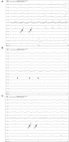
Three sets of consecutive EEG epochs in a 12 year old Shepherd mix illustrating evolution of GPDs to a focal seizure, and spontaneous resolution. The dog has a history of nasal aspergillosis and recent cluster seizures. Referential montage HF 35 Hz, sensitivity 5 uV/mm. (A) GPDs (arrows) progressively become more polyphasic, then develop bursts (>4 phases, longer than 500 ms) (circle). (B) GPDs (arrows) are associated with progressively longer bursts (circles), until a clinical focal seizure involving the left eye and face occurs, lasting 40 s. No pharmacologic intervention was given. Arrowheads indicate the start of electromyography (EMG) artifact from the muscle activity on the left frontal (F3, Fz) and left eye channels. (C) After termination of the clinical seizure, the GPDs (arrows) are apparent at a 2–2.5 Hz frequency, until they terminate spontaneously.

References

    1. Foreman B, Claassen J, Khaled KA, Jirsch J, Alschuler DM, Wittman J, et al. . Generalized periodic discharges in the critically ill: a case-control study of 200 patients. Neurology. (2012) 79:951–60. 10.1212/WNL.0b013e3182735cd7 - DOI - PMC - PubMed
    1. Zafar SF, Subramaniam T, Osman G, Herlopian A, Struck AF. Electrographic seizures and ictal-interictal continuum (IIC) patterns in critically ill patients. Epilepsy Behav. (2020) 106:107037. 10.1016/j.yebeh.2020.107037 - DOI - PubMed
    1. Chatrian GE, Shaw C-M, Leffman H. The significance of periodic lateralized epileptiform discharges in EEG: an electrographic, clinical and pathological study. Electroenceph Clin Neurophysiol. (1964) 17:177–93. 10.1016/0013-4694(64)90149-X - DOI - PubMed
    1. Chatrian GE. An EEG pattern: pseudo-rhythmic recurrent sharp waves. Its relationships to local cerebral anoxia and hypoxia. Electroenceph Clin Neurophysiol. (1961) 13:144.
    1. Witsch J, Frey H-P, Schmidt JM, Velazquez A, Falo CM, Reznik M, et al. . Electroencephalographic periodic discharges and frequency-dependent brain tissue hypoxia in acute brain injury. JAMA Neurol. (2017) 74:301–9. 10.1001/jamaneurol.2016.5325 - DOI - PMC - PubMed