. 2022 Oct;5(10):912-922.
doi: 10.1038/s41929-022-00848-6. Epub 2022 Oct 6.

Principles of target DNA cleavage and the role of Mg2+ in the catalysis of CRISPR-Cas9

Affiliations

Principles of target DNA cleavage and the role of Mg2+ in the catalysis of CRISPR-Cas9

Łukasz Nierzwicki et al. Nat Catal. 2022 Oct.

Abstract

At the core of the CRISPR-Cas9 genome-editing technology, the endonuclease Cas9 introduces site-specific breaks in DNA. However, precise mechanistic information to ameliorating Cas9 function is still missing. Here, multi-microsecond molecular dynamics, free-energy and multiscale simulations are combined with solution NMR and DNA cleavage experiments to resolve the catalytic mechanism of target DNA cleavage. We show that the conformation of an active HNH nuclease is tightly dependent on the catalytic Mg2+, unveiling its cardinal structural role. This activated Mg2+-bound HNH is consistently described through molecular simulations, solution NMR and DNA cleavage assays, revealing also that the protonation state of the catalytic H840 is strongly affected by active site mutations. Finally, ab-initio QM(DFT)/MM simulations and metadynamics establish the catalytic mechanism, showing that the catalysis is activated by H840 and completed by K866, rationalising DNA cleavage experiments. This information is critical to enhance the enzymatic function of CRISPR-Cas9 toward improved genome-editing.


Figures

Fig. 1.
Fig. 1.. Overview of the Streptococcus pyogenes CRISPR-Cas9 system.
(A) X-ray structure of the CRISPR-Cas9 system (PDB: 5F9R). The Cas9 protein is shown as ribbons, highlighting its catalytic domains HNH (green) and RuvC (blue), in complex with RNA (magenta) and DNA (black). (B) Close-up view on the HNH catalytic site, displaying the D839 and D831 residues coordinating Mg2+ and forming a catalytic triad with H840. This configuration of the catalytic core – referred as pseudo-active – arises from structures capturing HNH in the absence of Mg2+ (e.g., PDB: 5F9R) and from the homology with the T4 endonuclease VII (Supplementary Fig. 1). (C) Catalytic core from the cryo-EM structure EMD-0584 (PDB: 6O0Y) capturing HNH in the presence of Mg2+ after target strand cleavage, and (D) model of the catalytic site prior DNA cleavage. In this configuration – referred as active – N863 coordinates Mg2+ in place of D861. The atomic coordinates of HNH are shown as cartoon (green), while the electronic density is shown as wireframes (grey). The EMD-0584 map displays a visible density in the position of Mg2+ (C, indicated using an arrow), in agreement with the EMD-24838 map (Supplementary Fig. 2), enabling to locate the Mg2+ ion in the catalytic state (D).
Fig. 2.
Fig. 2.. Transition of the HNH domain from pseudo-active to active states.
(A) Free energy profiles for the HNH conformational transition in the presence of Mg2+ (w Mg, green) and without Mg2+ ions (w/o Mg, magenta). Two close-up views show the conformation of the pseudo-active (i.e., pseudo) and active states at their energetic minima. The HNH catalytic site bound to Mg2+ is shown as in Fig. 1C. Residues in green refer to HNH bound to Mg2+, while residues in magenta show the conformational change occurring in the absence of Mg2+ (also indicated using an arrow). (B) Probability distributions of critical interaction distances in the presence of Mg2+ (green) and absence of Mg2+ (magenta) for the pseudo-active and active states at their energetic minima (i.e., at −2.5 Å ≤ RC ≤ −1.5 Å and 1.5 Å ≤ RC ≤ 2.5 Å, respectively). The Welch’s t-test was used to assess the statistical significance of the differences in the distributions of the interaction distances with and without Mg2+. At the confidence level of 95 %, we rejected the null hypothesis in favour of the alternative with the p value < 0.0001. This observation was true for all cases but for the K866–D839 distance in the active system, where p = 0.437, as arising from overlapping distributions.
Fig. 3.
Fig. 3.. Chemical environment enabling the catalysis.
(A) Titration of the H799 and H840 side chains in the wild-type HNH (WT, top) and in the D893A mutant (bottom), reporting changes in the 1H-13Cε1 correlation as the histidine nitrogen atoms change protonation state, in the presence of Mg2+ and DNA. Resonances are coloured according to the pH values in the legend. Chemical shifts ~8.5 ppm correspond to fully protonated histidine, while those ~8.2–7.9 ppm refer to partially protonated or deprotonated residues. The trajectories of the chemical shifts are indicated using dashed arrows. In the D893A mutant, H840 shows evidence of two-three conformational states (upper arrow, H8401-3). (B) Fitted titration curves for pKa determination of H799 and H840 in the WT HNH through solution NMR (experimental) and constant pH molecular dynamics (theoretical). A modified version of the Henderson-Hasselbach equation was used for fitting the experimental data. Computational data were obtained by fitting the deprotonated fraction to Equation 3. Each data point represents the ensemble population of the protonated/deprotonated states from constant pH MD simulation (last ~32 ns) for each pH value. pKa values are reported, alongside the error from the fit (see Supplementary Methods). (C) Active state of HNH displaying the tautomeric form of H840 protonated on ε (H840-ε), which occurs for >60 % of CpH MD at pH 7.4 (Supplementary Fig. 11). (D) Titration curves for the catalytic H840 in the WT HNH and its mutants and change in pKa upon mutation (ΔpKa). In plots of the former, error associated with variance in NMR chemical shifts is smaller than the data points depicting Δδ. Values of ΔpKa are depicted as dots with error bars propagated as E(ΔpKa) = E(mutant) – E(wt), where E is the error determined from fits of the individual titration curves in Prism 9.0, centred at the calculated ΔpKa value. Coloured bars are shown as a visual aide to assess the magnitude of ΔpKa. (E) In vitro cleavage kinetics of Cas9 HNH mutants on a double stranded DNA on-target substrate. Line represents a single exponential fit of each individual time course experiment, each data point represents an average of four independent experiments (n = 4) with standard deviation plotted for each data point.
Fig. 4.
Fig. 4.. Effect of alanine mutations on the catalytic site.
Data are shown for the D839A, N863A and K866A mutants in the active state of HNH. Alanine mutations are shown in violet. (A) D839A affects the conformation of H840, resulting in three main conformations (H8401-3). (B) N863A results in the detachment of the S860–K866 α-helix from the catalytic Mg2+, destabilising the catalytic core. (C) K866A destabilises the catalytic core, with flexibility of the S860–D868 loop (four configurations are shown). Bottom graphs: stability of the catalytic site, computed as probability distribution of the heavy atoms’ Root Mean Square Deviation (RMSD) within 8 Å of the catalytic Mg2+; and location of the catalytic H840 with respect to the scissile phosphate (PSCI), computed considering the interatomic distance between H840 (Nδ) and PSCI. Data are reported for three simulation replicas of ~1 μs each. Vertical dashed lines (orange) indicate the cutoffs for the stability of the catalytic site (i.e., RMSD < 4 Å) and for the catalytic function of H840 (i.e., H840–PSCI < 6 Å allows the water nucleophile to position between H840 and PSCI, Fig. 3C, while H840–PSCI > 6 Å results in the detachment of H840 from the catalytic centre).
Fig. 5.
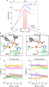
Fig. 5.. Free energy profiles for phosphodiester bond cleavage.
(A) Free energy profiles (ΔF, in kcal/mol) for the active (red) and pseudo-active (blue) states of HNH, obtained through QM(BLYP)/MM MD and Thermodynamic Integration. The difference in distance between the breaking and forming P–O bonds is the reaction coordinate (RC = d1–d2, shown in panel B). The chemical step evolves from the Reactants (R) to Products (P) passing through a Transition State (TS, region indicated using a red vertical bar). Error bars show standard deviations obtained from error propagation analysis of the primary data set in which each data point represents the mean from the last ~5 ps of converged ab-initio MD. (B) Close-up view of the TS structure in the active (left) and pseudo-active (right) states of HNH. (C) Dynamical RESP (D-RESP) charges within the TS region (i.e., −0.2 Å < RC < 0.2 Å) for the active and pseudo-active states. At each value of the RC, data are presented as mean (solid line) and standard deviation (shaded bands), computed over the last ~5 ps of converged ab-initio MD.
Fig. 6.
Fig. 6.. Catalytic mechanism of DNA cleavage in the HNH domain of CRISPR-Cas9.
Two-dimensional free energy surface for phosphodiester bond cleavage reporting the progress of the chemical step from the reactants (R), transition state (TS) and products (P) along two collective variables (CVs, shown on the 3D structure of the R). CV1 denotes the nucleophilic attack on scissile phosphate, while CV2 accounts for the proton transfer from the water nucleophile to H840. The free energy surface was obtained through QM/MM metadynamics. Unbiased QM/MM simulations of the P state reveal that the K866 side chain releases a proton to the water molecule coordinating Mg2+, which protonates the DNA O3’, leading to the final product (PFIN) of DNA cleavage. This clarifies DNA cleavage experiments (Fig. 3E), showing that the K866A substitution remarkably reduces the enzymatic activity.

Similar articles

Cited by

References

    1. Doudna JA & Charpentier E Genome editing. The new frontier of genome engineering with CRISPR-Cas9. Science 346, 1258096 (2014). - PubMed
    1. Jinek M et al. A Programmable Dual-RNA-Guided DNA Endonuclease in Adaptive Bacterial Immunity. Science 337, 816–821 (2012). - PMC - PubMed
    1. Jinek M et al. Structures of Cas9 Endonucleases Reveal RNA-Mediated Conformational Activation. Science 343, 1247997 (2014). - PMC - PubMed
    1. Nishimasu H et al. Crystal Structure of Cas9 in Complex with Guide RNA and Target DNA. Cell 156, 935–949 (2014). - PMC - PubMed
    1. Casalino L, Nierzwicki Ł, Jinek M & Palermo G Catalytic Mechanism of Non-Target DNA Cleavage in CRISPR-Cas9 Revealed by Ab Initio Molecular Dynamics. ACS Catal. 10, 13596–13605 (2020). - PMC - PubMed