. 2023 Feb 3:2023:4365895.
doi: 10.1155/2023/4365895. eCollection 2023.

Diabetes-Induced Autophagy Dysregulation Engenders Testicular Impairment via Oxidative Stress

Affiliations

Diabetes-Induced Autophagy Dysregulation Engenders Testicular Impairment via Oxidative Stress

Renfeng Xu et al. Oxid Med Cell Longev. .

Abstract

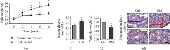
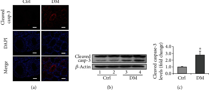
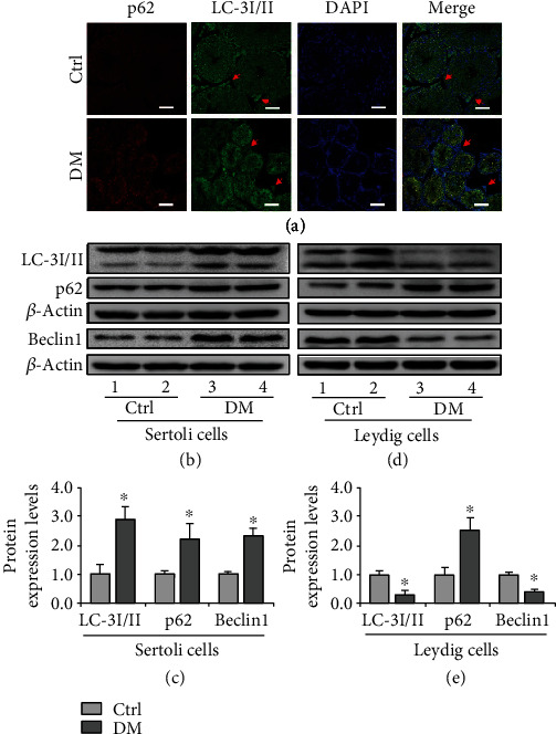
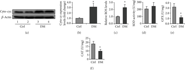
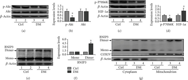
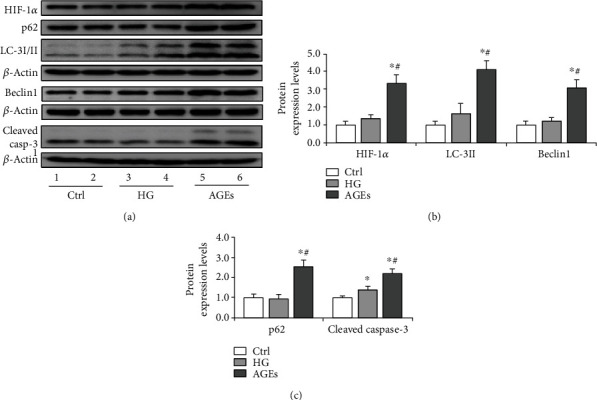
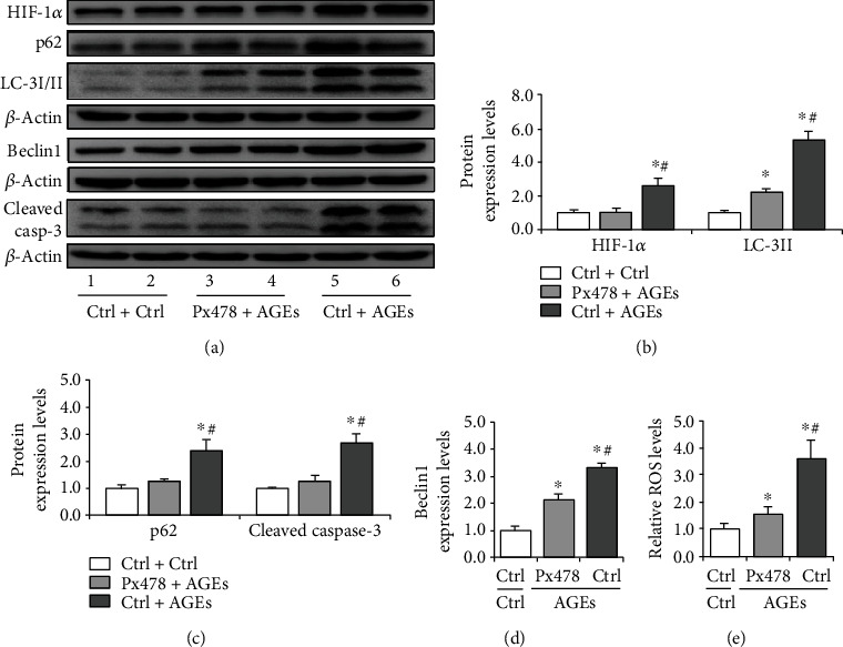
Testes produce sperms, and gamete generation relies on a proper niche environment. The disruption of hierarchical regulatory homeostasis in Leydig or Sertoli cells may evoke a sterile phenotype in humans. In this study, we recapitulated type 2 diabetes mellitus by using a high-fat diet- (HFD-) fed mouse model to identify the phenotype and potential mechanism of diabetes-induced testicular impairment. At the end of the study, blood glucose levels, testosterone structure, testicular antioxidant capacity, and testosterone level and the expression of hypoxia-inducible factor- (HIF-) 1α, apoptosis-related protein cleaved-caspase3, and autophagy-related proteins such as LC3I/II, p62, and Beclin1 were evaluated. We found that long-term HFD treatment causes the development of diabetes mellitus, implicating increased serum glucose level, cell apoptosis, and testicular atrophy (P < 0.05 vs. Ctrl). Mechanistically, the results showed enhanced expression of HIF-1α in both Sertoli and Leydig cells (P < 0.05 vs. Ctrl). Advanced glycation end products (AGEs) were demonstrated to be a potential factor leading to HIF-1α upregulation in both cell types. In Sertoli cells, high glucose treatment had minor effects on Sertoli cell autophagy. However, AGE treatment stagnated the autophagy flux and escalated cell apoptosis (P < 0.05 vs. Ctrl+Ctrl). In Leydig cells, high glucose treatment was adequate to encumber autophagy induction and enhance oxidative stress. Similarly, AGE treatment facilitated HIF-1α expression and hampered testosterone production (P < 0.05 vs. Ctrl+Ctrl). Overall, these findings highlight the dual effects of diabetes on autophagy regulation in Sertoli and Leydig cells while imposing oxidative stress in both cell types. Furthermore, the upregulation of HIF-1α, which could be triggered by AGE treatment, may negatively affect both cell types. Together, these findings will help us further understand the molecular mechanism of diabetes-induced autophagy dysregulation and testicular impairment, enriching the content of male reproductive biology in diabetic patients.


Conflict of interest statement

The authors declare that they have no competing interests.

Figures

Figure 1
Figure 1
Changes of testicular weight and histology in diabetic mice induced by high-fat diet. (a) The growth curve of mice fed with normal control diet or high-fat diet. (b) Effect of diabetes on fasting glucose. (c) Effect of diabetes on testicular weight. (d) Effect of diabetes on testicular histology. The testicular morphology of diabetic mice was impaired, with disordered arrangement of spermatocytes observed by H&E staining. DM: diabetes mellitus. P < 0.05 was considered to indicate a statistically significant difference. Bar = 100 μm. P < 0.05 vs. Ctrl.
Figure 2
Figure 2
Expression of cell apoptosis marker, cleaved caspase-3, in the testis of diabetic mice. (a) The immunofluorescent staining of cleaved caspase-3 in the mouse testis. (b) Representative immunoblotting of cleaved caspase-3. (c) Densitometric qualification of cleaved caspase-3. DM: diabetes mellitus. P < 0.05 was considered to indicate a statistically significant difference. Bar = 100 μm. P < 0.05 vs. Ctrl.
Figure 3
Figure 3
Expression of LC-3I/II, p62, and Beclin1 in the testicular cells in vivo and in vitro. (a) The immunofluorescent staining of p62 and LC-3I/II in the testis of diabetic mice. Red arrow indicates Leydig cells. (b) Representative immunoblotting of LC-3I/II, p62, and Beclin1 in Sertoli cells. (c) Densitometric qualification of LC-3II, p62, and Beclin1 in Sertoli cells. (d) Representative immunoblotting of LC-3I/II, p62, and Beclin1 in Leydig cells. (e) Densitometric quantification of LC-3II, p62, and Beclin1 in Leydig cells. Diabetes exerts dual effects on autophagy regulation in Sertoli and Leydig cells. DM: diabetes mellitus. P < 0.05 was considered to indicate a statistically significant difference. Bar = 100 μm. P < 0.05 vs. Ctrl.
Figure 4
Figure 4
Expression of oxidative stress-related marker proteins in the diabetic testis and Leydig cells. (a) Representative immunoblotting of Cyto-cyc in the testis of diabetic mice. (b) Densitometric qualification of Cyto-cyc in the testis of diabetic mice. (c) Relative ROS levels in the testis of diabetic mice. (d) SOD activity in the testis of diabetic mice. (e) GPX content in the testis of diabetic mice. (f) CAT content in the testis of diabetic mice. DM: diabetes mellitus; Rap: rapamycin; HG: high glucose. P < 0.05 was considered to indicate a statistically significant difference. P < 0.05 vs. Ctrl.
Figure 5
Figure 5
Expression of Cyto-cyc and LC-3 proteins in the diabetic testis and Leydig cells. (a) Representative immunoblotting of Cyto-cyc and LC-3I/II in Leydig cells treated with Rap and HG. (b) Densitometric qualification of Cyto-cyc in Leydig cells treated with Rap and HG. (c) Densitometric qualification of LC-3II in Leydig cells treated with Rap and HG. (d) Relative ROS levels in Leydig cells treated with Rap and HG. DM: diabetes mellitus; Rap: rapamycin; HG: high glucose. P < 0.05 was considered to indicate a statistically significant difference. #P < 0.05 vs. Ctrl+Ctrl. &P < 0.05 vs. Rap+HG.
Figure 6
Figure 6
Activation of HIF-1α pathway in Sertoli cells of diabetic mice. (a) Representative immunoblotting of Akt and p-Akt in the testis of diabetic mice. (b) Densitometric qualification of Akt and p-Akt in the testis of diabetic mice. (c) Representative immunoblotting of p-P70S6K and HIF-1α in the testis of diabetic mice. (d) Densitometric qualification of p-P70S6K and HIF-1α in the testis of diabetic mice. (e) Representative immunoblotting of dimer and monomer BNIP3 in the testis of diabetic mice. (f) Densitometric qualification of dimer and monomer BNIP3 in the testis of diabetic mice. (g) Representative immunoblotting of dimer and monomer BNIP3 in the cytoplasm and mitochondrion. DM: diabetes mellitus. P < 0.05 was considered to indicate a statistically significant difference. P < 0.05 vs. Ctrl.
Figure 7
Figure 7
Expression of HIF-1α, LC-3I/II, p62, and cleaved caspase-3 during AGE-induced oxidative stress in Sertoli cells. Sertoli cells were treated with HG (20 mM) or AGEs (100 μg/ml) for 72 h. AGEs aggravated HIF-1α expression and induced autophagy dysregulation. (a) Representative immunoblotting of HIF-1α, LC-3I/II, p62, Beclin1, and cleaved caspase-3 in Sertoli cells treated with HG and AGEs. (b) Densitometric qualification of HIF-1α, LC-3II, and Beclin1 in Sertoli cells treated with HG and AGEs. (c) Densitometric qualification of p62 and cleaved caspase-3 in Sertoli cells treated with HG and AGEs. P < 0.05 was considered to indicate a statistically significant difference. P < 0.05 vs. Ctrl. #P < 0.05 vs. HG.
Figure 8
Figure 8
Effect of Px478 on HIF-1α, LC-3I/II, p62, and cleaved caspase-3 expression during AGE-induced oxidative stress in Sertoli cells. The cells were treated with AGEs (100 μg/ml) for 72 h, and Px478 (20 μM) was added 16 h before harvesting. (a) Representative immunoblotting of HIF-1α, LC-3I/II, p62, Beclin1, and cleaved caspase-3 in Sertoli cells treated with Px478 and AGEs. (b) Densitometric qualification of HIF-1α, LC-3II, and Beclin1 in Sertoli cells treated with Px478 and AGEs. (c) Densitometric qualification of p62 and cleaved caspase-3 in Sertoli cells treated with Px478 and AGEs. (d) Relative ROS levels in Sertoli cells treated with Px478 and AGEs. P < 0.05 was considered to indicate a statistically significant difference. P < 0.05, vs. Ctrl+Ctrl. #P < 0.05, vs. Px478+AGEs.
Figure 9
Figure 9
Expression of HIF-1α in the diabetic testis and Leydig cells. Leydig cells were treated with AGEs (100 μg/ml) for 72 h, and Px478 was added 16 h before harvesting. (a) Representative immunoblotting of HIF-1α in the testis of diabetic mice. (b) Densitometric qualification of HIF-1α in the testis of diabetic mice. (c) Serum testosterone level in diabetic mice. (d) Representative immunoblotting of HIF-1α in Leydig cells treated with Px478 and AGEs. (e) Densitometric qualification of HIF-1α in Leydig cells treated with Px478 and AGEs. (f) Testosterone level in supernatant of Leydig cells treated with Px478 and AGEs. P < 0.05 was considered to indicate a statistically significant difference. P < 0.05 vs. Ctrl. #P < 0.05 vs. Ctrl+Ctrl. &P < 0.05, vs. Px478+AGEs.

Similar articles

Cited by

References

    1. Claman P. Men at risk: occupation and male infertility. Fertility and Sterility . 2004;81:19–26. doi: 10.1016/S0015-0282(03)01188-9. - DOI - PubMed
    1. Sharpe R. M., Franks S. Environment, lifestyle and infertility -- an inter-generational issue. Nature Medicine . 2002;8(S10):S33–S40. doi: 10.1038/nm-fertilityS33. - DOI - PubMed
    1. Semet M., Paci M., Saïas-Magnan J., et al. The impact of drugs on male fertility: a review. Andrology . 2017;5(4):640–663. doi: 10.1111/andr.12366. - DOI - PubMed
    1. Homan G., Davies M., Norman R. The impact of lifestyle factors on reproductive performance in the general population and those undergoing infertility treatment: a review. Human Reproduction Update . 2007;13(3):209–223. doi: 10.1093/humupd/dml056. - DOI - PubMed
    1. Hayden R. P., Flannigan R., Schlegel P. N. The role of lifestyle in male infertility: diet, physical activity, and body habitus. Current Urology Reports . 2018;19(7):p. 56. doi: 10.1007/s11934-018-0805-0. - DOI - PubMed