. 2023 Feb 2:2023:1493684.
doi: 10.1155/2023/1493684. eCollection 2023.

Simvastatin Reduces NETosis to Attenuate Severe Asthma by Inhibiting PAD4 Expression

Affiliations

Simvastatin Reduces NETosis to Attenuate Severe Asthma by Inhibiting PAD4 Expression

Yun-Rong Chen et al. Oxid Med Cell Longev. .

Abstract

Objective: Patients with severe asthma respond poorly to corticosteroids, and their care accounts for more than 60% of the total costs attributed to asthma. Neutrophils form neutrophil extracellular traps (NETs), which play a crucial role in severe asthma. Statins have shown anti-inflammatory effects by reducing NETosis. In this study, we investigate if simvastatin can attenuate severe asthma by reducing NETosis and the underlying mechanism.

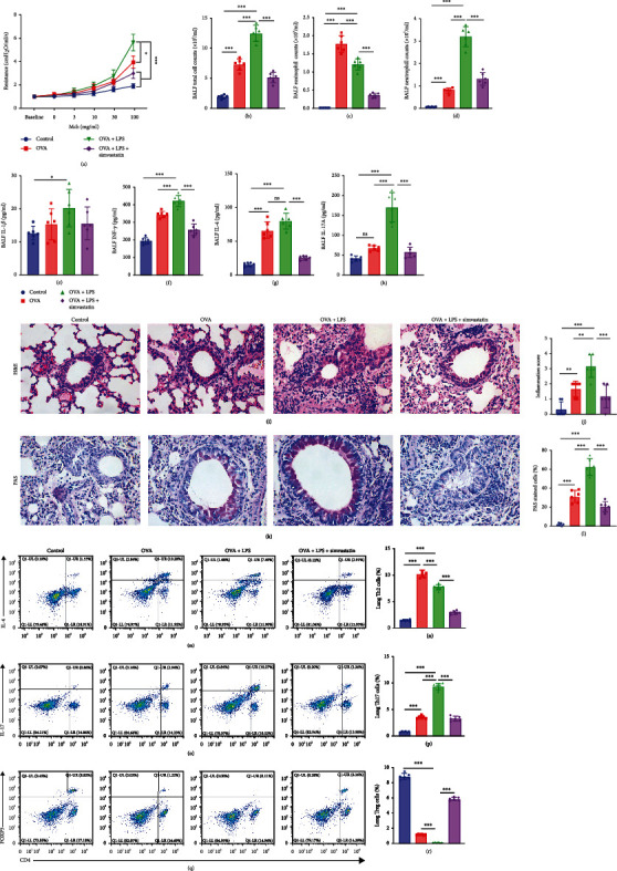
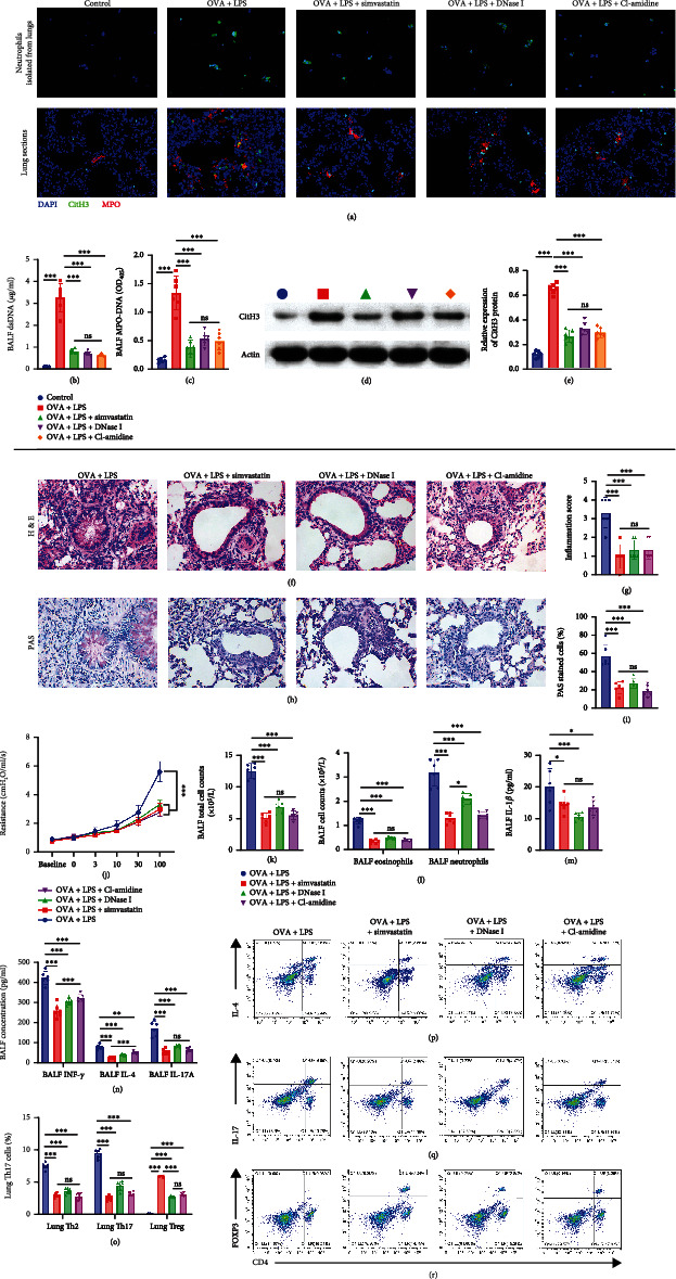
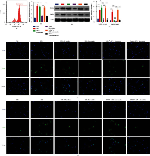
Methods: Mice were concomitantly sensitized with ovalbumin (OVA), house dust mite (HDM), and lipopolysaccharide (LPS) during sensitization to establish a mouse model of severe asthma with neutrophil predominant inflammation (OVA+LPS mice) and treated with or without simvastatin. In inflammatory response, proportions of Th2, Th17, and Treg cells in lung tissue were detected by flow cytometry, and the levels of cytokines, dsDNA, and MPO-DNA in bronchoalveolar lavage fluid (BALF) were analyzed by ELISA. Citrullinated histone H3 (CitH3) and peptidyl arginine deiminase 4 (PAD4) in lung tissue were determined by Western blot and immunofluorescence imaging. PAD4 mRNA was determined by quantitative PCR (qPCR). HL-60 cells were differentiated into neutrophil-like cells by 1.25% DMSO. The neutrophil-like cells were treated with or without LPS, and simvastatin was then stimulated with PMA. CitH3 and PAD4 expressions were determined.

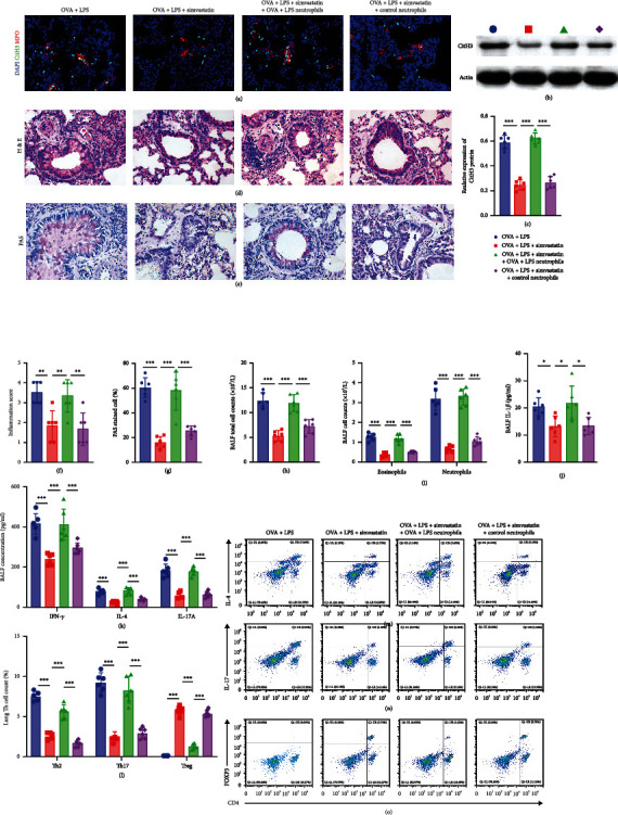
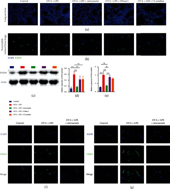
Results: Sensitization with OVA, HDM, and LPS resulted in neutrophilic inflammation and the formation of NETs in the lungs. Simvastatin treatment reduced the inflammation score, cytokine levels, total cells, and neutrophil counts in the BALF and reduced proportions of Th2 and Th17 but increased Treg cells in lungs of OVA+LPS mice. Simvastatin-treated OVA+LPS mice show reduced NET formation in BALF and lung tissue compared to control mice. Adoptive transfer of neutrophils was sufficient to restore NETosis and neutrophilic inflammation in simvastatin-treated OVA+LPS mice. Simvastatin reduced PAD4 mRNA and protein expression in lung tissues and neutrophils isolated from lungs of OVA+LPS mice and consequent NET formation. In vitro, simvastatin reduced LPS-induced PAD4 upregulation and NETosis in HL-60-differentiated neutrophil-like cells. Furthermore, PAD4-overexpressed lentiviral transduction was sufficient to restore PAD4 protein expression and NETosis in simvastatin-treated HL-60-differentiated neutrophil-like cells.

Conclusions: Simvastatin reduces Th17-mediated neutrophilic inflammation and airway hyperreactivity by reducing PAD4 expression and inhibiting NETosis in a mouse model of severe asthma. Severe asthmatic patients with high levels of circulating NETs or sputum NETs may show improved responses to statin treatment.


Conflict of interest statement

The authors declare that the research was conducted in the absence of any commercial or financial relationships that could be construed as a potential conflict of interest.

Figures

Figure 1
Figure 1
Simvastatin treatment ameliorates features of TH2-mediated and TH17-mediated asthma in mice. (a) Measurement of dynamic airway resistance. (b–d) Total cell counts, eosinophil counts, and neutrophil counts (105 cells/ml) in the BALF. (e–h) Levels of IL-1β, IFN-γ, IL-4, and IL-17 in the BALF. (i, j) Representative hematoxylin and eosin (H&E) staining and inflammation score of lung sections of mice. (k, l) Representative PAS staining of lung sections and quantification of PAS-stained epithelial cells per bronchi showing airway mucus production in mice. (m–r) Flow cytometric analysis of percentages of TH2, TH17, and Treg lymphocytes in the lung.
Figure 2
Figure 2
Simvastatin treatment attenuates TH17-mediated neutrophilic asthma through reducing NETosis in mouse lungs. (a) Confocal microscopy staining of CitH3+ MPO+ DAPI+ NETs released from ex vivo-cultured neutrophils and the lung sections of the indicated groups of mice. (b) Levels of extracellular dsDNA in the BALF. (c) ELISA measurement of MPO-DNA complexes in the BALF of mice. (d) Representative blots of CitH3 and actin (loading control) assessed by Western blot of lung protein extracts from mice. (e) Quantification of normalized CitH3 levels in lung protein extracts of mice. (f, g) Representative hematoxylin and eosin (H&E) staining and inflammation score of lung sections of mice. (h) Representative PAS staining of lung sections of mice. (i) Quantification of PAS-stained epithelial cells per bronchi showing airway mucus production in mice. (j) Measurement of dynamic airway resistance. (k, l) Total cell counts, eosinophil counts, and neutrophil counts (105 cells/ml) in the BALF. (m, n) Levels of IL-1β, IFN-γ, IL-4, and IL-17A in the BALF. (o–r) Flow cytometric analysis of percentages of TH2, TH17, and Treg lymphocytes in the lung.
Figure 3
Figure 3
Adoptively transferring neutrophils from OVA+LPS mice to simvastatin-treated OVA+LPS mice was sufficient to trigger the TH17-mediated neutrophilic asthma. (a) Confocal microscopy staining of CitH3+ MPO+ DAPI+ NETs released from ex vivo-cultured neutrophils and the lung sections of the indicated groups of mice. (b) Representative blots of CitH3 and actin (loading control) assessed by Western blot of lung protein extracts from mice. (c) Quantification of normalized CitH3 levels in lung protein extracts of mice. (d, f) Representative hematoxylin and eosin (H&E) staining of lung sections of mice. (e) Representative PAS staining of lung sections of mice. (g) Quantification of PAS-stained epithelial cells per bronchi showing airway mucus production in mice. (h, i) Total cell counts, eosinophil counts, and neutrophil counts (105 cells/ml) in the BALF. (j, k) Levels of IL-1β, IFN-γ, IL-4, and IL-17 in the BALF. (l–o) Flow cytometric analysis of percentages of TH2, TH17, and Treg lymphocytes in the lung.
Figure 4
Figure 4
Simvastatin treatment reduces NETosis via PAD4 in the lungs of OVA+LPS mice. (a, b) Representative immunofluorescence images of PAD4 and DAPI staining of lung sections (n = 6) and ex vivo-cultured neutrophils in the indicated groups of mice. (c) Representative blots of PAD4 and actin (loading control) assessed by Western blot of lung protein extracts from mice. (d) Quantification of normalized PAD4 levels in lung protein extracts of mice. (e) The expression levels of PAD4 mRNA examined by RT-qPCR in lung homogenates of mice. (f, g) The lung neutrophils were isolated from OVA+LPS mice or control mice and preincubated the neutrophils in media with or without simvastatin (10 μg/ml) before stimulation with 100 nmol/l phorbol 12-myristate 13-acetate (PMA). (f) Representative immunofluorescence images of PAD4 and DAPI staining. (g) Representative immunofluorescence images of CitH3 and DAPI staining.
Figure 5
Figure 5
Simvastatin treatment reduces NET formation in neutrophils via PAD4 in vitro. (a) Flow cytometry analysis of CD11b expression on HL-60 cells after 5 days of culture with DMSO. (b) The expression levels of PAD4 mRNA examined by RT-qPCR in the HL-60-differentiated neutrophil-like cells. (c) Representative blots of PAD4 and actin (loading control) assessed by Western blot of the HL-60-differentiated neutrophil-like cells. (d) Quantification of normalized PAD4 levels in the HL-60-differentiated neutrophil-like cells. (e) Representative immunofluorescence images of PAD4 and DAPI staining in the HL-60-differentiated neutrophil-like cells. (f) Representative immunofluorescence images of CitH3 and DAPI staining in the HL-60-differentiated neutrophil-like cells.

References

    1. Schoettler N., Strek M. E. Recent advances in severe asthma: from phenotypes to personalized medicine. Chest . 2020;157(3):516–528. doi: 10.1016/j.chest.2019.10.009. - DOI - PMC - PubMed
    1. Israel E., Reddel H. K. Severe and difficult-to-treat asthma in adults. The New England Journal of Medicine . 2017;377(10):965–976. doi: 10.1056/NEJMra1608969. - DOI - PubMed
    1. Holgate S. T. Innate and adaptive immune responses in asthma. Nature Medicine . 2012;18(5):673–683. doi: 10.1038/nm.2731. - DOI - PubMed
    1. Wang Y. H., Wills-Karp M. The potential role of interleukin-17 in severe asthma. Current Allergy and Asthma Reports . 2011;11(5):388–394. doi: 10.1007/s11882-011-0210-y. - DOI - PMC - PubMed
    1. Krishnamoorthy N., Douda D. N., Brüggemann T. R., et al. Neutrophil cytoplasts induce TH17 differentiation and skew inflammation toward neutrophilia in severe asthma. Science Immunology . 2018;3(26, article eaao4747) doi: 10.1126/sciimmunol.aao4747. - DOI - PMC - PubMed