

This is a preprint.
Altered transcriptomes, cell type proportions, and dendritic spine morphology in hippocampus of suicide deaths
- PMID: 36778310
- PMCID: PMC9915834
- DOI: 10.1101/2023.01.28.23285121
Altered transcriptomes, cell type proportions, and dendritic spine morphology in hippocampus of suicide deaths
Update in
-
Altered transcriptomes, cell type proportions, and dendritic spine morphology in hippocampus of suicide decedents.J Affect Disord. 2024 Dec 15;367:118-128. doi: 10.1016/j.jad.2024.08.144. Epub 2024 Aug 25. J Affect Disord. 2024. PMID: 39191313
Abstract
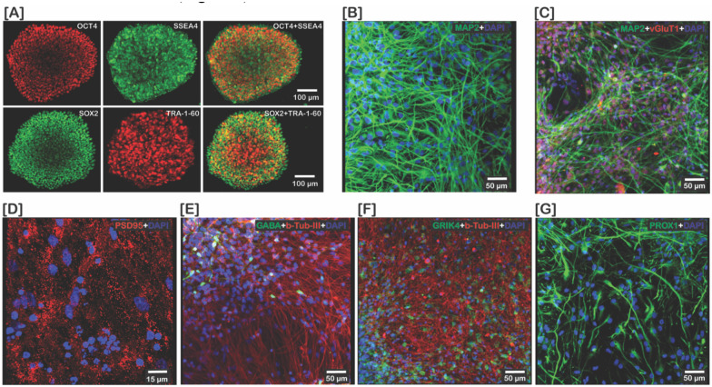
Suicide is a condition resulting from complex environmental and genetic risks that affect millions of people globally. Both structural and functional studies identified the hippocampus as one of the vulnerable brain regions contributing to suicide risk. Here, we have identified the hippocampal transcriptomes, gene ontology, cell type proportions, dendritic spine morphology, and transcriptomic signature in iPSC-derived neuronal precursor cells (NPCs) and neurons in postmortem brain tissue from suicide deaths. The hippocampal tissue transcriptomic data revealed that NPAS4 gene expression was downregulated while ALDH1A2, NAAA, and MLXIPL gene expressions were upregulated in tissue from suicide deaths. The gene ontology identified 29 significant pathways including NPAS4-associated gene ontology terms "excitatory post-synaptic potential", "regulation of postsynaptic membrane potential" and "long-term memory" indicating alteration of glutamatergic synapses in the hippocampus of suicide deaths. The cell type deconvolution identified decreased excitatory neuron proportion and an increased inhibitory neuron proportion providing evidence of excitation/inhibition imbalance in the hippocampus of suicide deaths. In addition, suicide deaths had increased dendric spine density, due to an increase of thin (relatively unstable) dendritic spines, compared to controls. The transcriptomes of iPSC-derived hippocampal-like NPCs and neurons revealed 31 and 33 differentially expressed genes in NPC and neurons, respectively, of suicide deaths. The suicide-associated differentially expressed genes in NPCs were RELN, CRH, EMX2, OXTR, PARM1 and IFITM2 which overlapped with previously published results. The previously-known suicide-associated differentially expressed genes in differentiated neurons were COL1A1, THBS1, IFITM2, AQP1, and NLRP2. Together, these findings would help better understand the hippocampal neurobiology of suicide for identifying therapeutic targets to prevent suicide.
Conflict of interest statement
Competing Interests The authors have nothing to disclose
Figures


References
-
- Sisti D, Mann JJ, Oquendo MA. Toward a Distinct Mental Disorder-Suicidal Behavior. JAMA Psychiatry. 2020;77(7):661–62. - PubMed
-
- Zhang L, Lucassen PJ, Salta E, Verhaert P, Swaab DF. Hippocampal neuropathology in suicide: Gaps in our knowledge and opportunities for a breakthrough. Neurosci Biobehav Rev. 2022;132:542–52. - PubMed
-
- Sinyor M, Williams M, Mitchell R, Zaheer R, Bryan CJ, Schaffer A, et al. Cognitive behavioral therapy for suicide prevention in youth admitted to hospital following an episode of self-harm: A pilot randomized controlled trial. J Affect Disord. 2020;266:686–94. - PubMed
Publication types
Grants and funding
LinkOut - more resources
Full Text Sources
Miscellaneous