[Preprint]. 2023 Feb 2:2023.02.02.526753.
doi: 10.1101/2023.02.02.526753.

Replicative Acinetobacter baumannii strains interfere with phagosomal maturation by modulating the vacuolar pH

Affiliations

Replicative Acinetobacter baumannii strains interfere with phagosomal maturation by modulating the vacuolar pH

Jesus S Distel et al. bioRxiv. .

Update in

Abstract

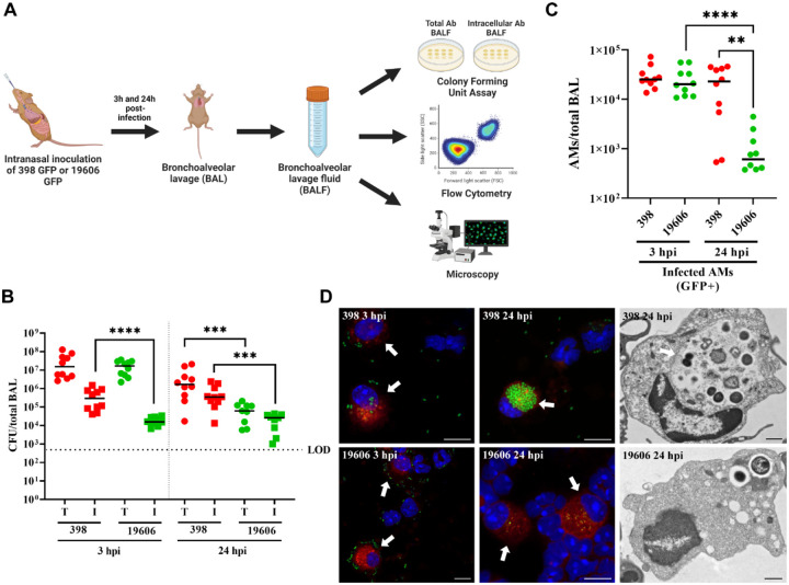
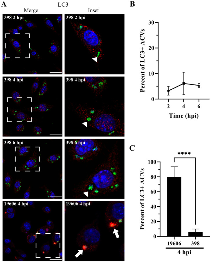
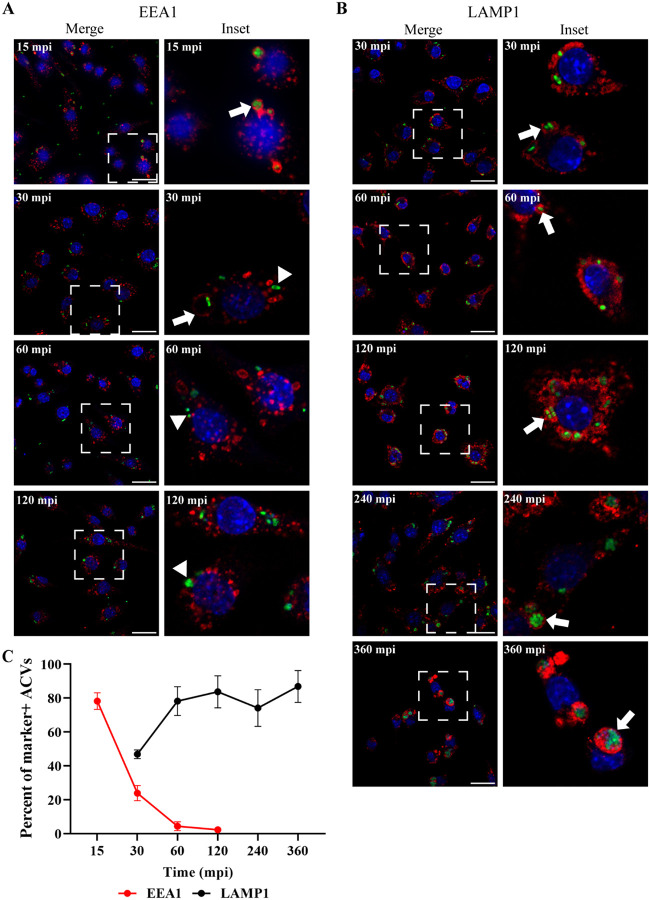
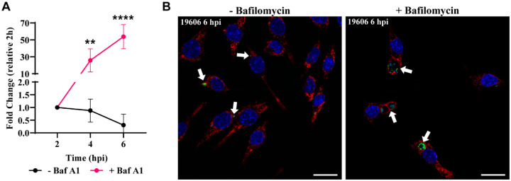
Bacterial pneumonia is a common infection of the lower respiratory tract that can afflict patients of all ages. Multidrug-resistant strains of Acinetobacter baumannii are increasingly responsible for causing nosocomial pneumonias, thus posing an urgent threat. Alveolar macrophages play a critical role in overcoming respiratory infections caused by this pathogen. Recently, we and others have shown that new clinical isolates of A. baumannii , but not the common lab strain ATCC 19606 (19606), can persist and replicate in macrophages within spacious vacuoles that we called A cinetobacter C ontaining V acuoles (ACV). In this work, we demonstrate that the modern A. baumannii clinical isolate 398, but not the lab strain 19606, can infect alveolar macrophages and produce ACVs in vivo in a murine pneumonia model. Both strains initially interact with the alveolar macrophage endocytic pathway, as indicated by EEA1 and LAMP1 markers; however, the fate of these strains diverges at a later stage. While 19606 is eliminated in an autophagy pathway, 398 replicates in ACVs and are not degraded. We show that 398 reverts the natural acidification of the phagosome by secreting large amounts of ammonia, a by-product of amino acid catabolism. We propose that this ability to survive within macrophages may be critical for the persistence of clinical A. baumannii isolates in the lung during a respiratory infection.


Conflict of interest statement

Competing Interest Statement: The authors declare no conflict of interest.

Figures

Figure 1.
Figure 1.. A. baumannii clinical isolate 398 infects AMs in vivo and survives inside ACVs.
(A) Schematic of the pneumonia model employed in this study. (B) Mice were intranasally infected with 1 × 10⁸ CFU of A. baumannii strains GFP-19606 or GFP-398. At 3 or 24 hpi the total (T) or intracellular (I) CFU from bronchoalveolar lavages were determined. Symbols represents individual animals. Median values are shown as horizontal black bars. Data from two independent experiments with 5 mice per experimental group are shown. (C) Quantification of infected AMs (CD45+CD11c+SiglecF+CD11b-GFP+) obtained from BALF at 3 hpi or 24 hpi with GFP-19606 or GFP-398 strains. Symbols represent individual animals. (D) Representative confocal microscopy images of cells present in the BALF of infected mice (left and central images). Cell nuclei were stained with DAPI (blue), A baumannii 19606 or 398 were detected by GFP fluorescence (green), and SiglecF was immunolabeled with specific antibodies (red). Arrows indicate AMs. Scale bars: 10 μm. Representative transmission electron microscopy images with infected AMs from BALF are shown at the right. Scale bars: 1 μm. Statistical analyses were performed using the Mann–Whitney test, **p < 0,0021, ****p < 0,0001. Hpi: hours post-infection.
Figure 2.
Figure 2.. 398 ACV does not colocalize with the autophagic marker LC3.
(A) Representative confocal microscopy micrographs of J774A.1 cells infected with A. baumannii strains GFP-398 or GFP-19606 at the different times post-infection (pi). Cell nuclei were stained with DAPI (blue), bacteria overexpress GFP (green) and LC3 was immunostained with a specific antibody (red). Scale bars: 20 μm. Insets (40 μm) are a higher magnification of the area indicated with a white box in the corresponding image. LC3-positive ACVs are indicated with arrows and LC3–negative ACVs are indicated with arrowheads. (B) Quantification of LC3 positive ACVs of the clinical isolate 398 at the different times pi. (C) Comparison of the percent of LC3 positive ACVs of 398 and 19606 at 4 hpi. At least 200 infected cells were analyzed per strain per time point. The results are expressed as mean ± standard error of the mean (SEM) of three independent experiments. Statistical analyses were performed using Welch’s t-test, **** < 0.0001.
Figure 3.
Figure 3.. The ACV interacts with the endocytic pathway.
J774A.1 cells were infected with A. baumannii GFP-398, and after the indicated times pi the cells were fixed and processed for confocal microscopy. The samples were stained to detect cell nuclei (blue), GFP-bacteria (green) and (A) EEA1 (red) or (B) LAMP1 (red). Bars: 20 μm. Insets (40 μm) are a higher magnification of the region indicated with a white box in the corresponding image. The presence of EEA1 or LAMP1 markers in the ACV is indicated by arrows while marker negative ACVs are denoted by arrowheads. (C) The percent of EEA1 or LAMP1 positive ACVs was determined at different times pi. At least 200 infected cells were analyzed per indicated time point. The results are expressed as mean ± SEM of three independent experiments. Mpi: minutes post-infection.
Figure 4.
Figure 4.. A. baumannii 398, but not 19606, resides in a non-degradative ACV.
(A) J774A.1 cells infected with A. baumannii 398 or 19606 were incubated with DQ-BSA and then fixed at the indicated time points. The samples were stained to detect cell nuclei (blue), DQ-BSA (green), A. baumannii (red) and actin (pink). Bars: 20 μm. Insets (20 μm) are a higher magnification of the area denoted in the corresponding image with a white box. (B) Fluorescent intensity plots of representative ACVs (indicated with arrows). (C) Quantification of DQ-BSA positive ACVs of the clinical isolate 398 at the different times pi. (D) Comparison of the percentage of DQ-BSA positive ACVs of 398 and 19606 at 4 hpi. Statistical analyses were performed using the Welch’s t-test, **** < 0.0001. At least 200 infected cells were analyzed per strain, per time point. Results are expressed as mean ± SEM of three independent experiments.
Figure 5.
Figure 5.. Bafilomycin A1 treatment allows 19606 to replicate in macrophages.
(A) J774A.1 macrophages were infected with GFP-19606 and treated with the proton pump V-ATPase inhibitor bafilomycin A1. Total numbers of intracellular CFU were determined at different times pi in treated and non-treated cells. Statistical analyses were performed by two-way ANOVA-test, ** < 0.0021, **** < 0.0001. (B) Representative images of cells infected with GFP-19606 (green) and incubated with or without bafilomycin A1 at 6 hpi are shown. Cell nuclei were stained with DAPI (blue) and LAMP1 with specific antibody (red). Bars: 20 μm.
Figure 6.
Figure 6.. 398 increases the luminal pH of the ACV during infection.
(A) J774A.1 cells infected with mCherry-398 (red) were incubated with LysoSensor (green) 15 minutes before the indicated time points. Samples were analyzed by in vivo confocal microscopy. Bars: 20 μm. (B) Analysis of the Mean Fluorescence Intensity (MFI) signal of LysoSensor per ACV at different times pi. (C) Percentage of ACVs that colocalize with LysoSensor at 2, 4, 6 and 24 hpi. Statistical analyses were performed using one way ANOVA-test, ** < 0.0021, *** < 0.0002. At least 200 infected cells were analyzed per indicated time point. Results are expressed as mean ± SEM of three independent experiments.
Figure 7.
Figure 7.. Intracellular replication correlates with the ability to grow at acidic pH.
(A) The intracellular replication of A. baumannii strains 398 and 19606 in J774A.1 macrophages was determined by antibiotic protection assays. (B) Growth curves of 398 and 19606 in buffered LB pH 5 as measured by OD600. (C) Changes in culture pH during A. baumannii growth, determined by phenol red absorbance at 560 nm. (D) Concentration of ammonia in LB cultures of 398 and 19606 strains at 4, 5 and 6 h post-inoculation. Results are expressed as mean ± SEM of three independent experiments.
Figure 8.
Figure 8.. Proposed model of the intracellular lifestyle of A. baumannii in macrophages.
Replicative strains of A. baumannii (green bacterium) interact with the endocytic pathway sequentially acquiring EEA1 and LAMP1 markers in the ACV. However, during its maturation, the ACV does not interact with autophagosomes or lysosomes of the host cell. Moreover, the replication of intracellular bacteria produces ammonia which neutralizes the luminal microenvironment of the ACV. On the other hand, non-replicative strains of A. baumannii (gray bacterium) are phagocytized by macrophages and reside in a nascent phagosome. This compartment maturates to a late phagosome, like the replicative ACV, by interactions with the endocytic pathway. Nevertheless, the final fate of the non-replicative strains of A. baumannii is the degradation in an autophagolysosome.

References

    1. Actis L. A., Smoot J. C., Barancin C. E., & Findlay R. H. (1999). Comparison of differential plating media and two chromatography techniques for the detection of histamine production in bacteria. Journal of Microbiological Methods, 39(1), 79–90. 10.1016/S0167-7012(99)00099-8 - DOI - PubMed
    1. Ambrosi C., Scribano D., Sarshar M., Zagaglia C., Singer B. B., & Palamara A. T. (2020). Acinetobacter baumannii Targets Human Carcinoembryonic Antigen-Related Cell Adhesion Molecules (CEACAMs) for Invasion of Pneumocytes. MSystems, 5(6), 1–17. 10.1128/mSystems.00604-20 - DOI - PMC - PubMed
    1. Banerjee S., & Kane P. M. (2020). Regulation of V-ATPase Activity and Organelle pH by Phosphatidylinositol Phosphate Lipids. Frontiers in Cell and Developmental Biology, 8(6). 10.3389/fcell.2020.00510 - DOI - PMC - PubMed
    1. Bissonnette E. Y., Lauzon-Joset J.-F., Debley J. S., & Ziegler S. F. (2020). Cross-Talk Between Alveolar Macrophages and Lung Epithelial Cells is Essential to Maintain Lung Homeostasis. Frontiers in Immunology, 11(10), 1–12. 10.3389/fimmu.2020.583042 - DOI - PMC - PubMed
    1. Brumell J. (2004). Salmonella redirects phagosomal maturation. Current Opinion in Microbiology, 7(1), 78–84. 10.1016/j.mib.2003.12.005 - DOI - PubMed

Publication types

LinkOut - more resources